7KD7 | pdb_00007kd7

Crystal structure of human NatD (NAA40) bound to a bisubstrate analogue


Experimental Data Snapshot

  • Method: X-RAY DIFFRACTION
  • Resolution: 1.44 Å
  • R-Value Free: 
    0.183 (Depositor), 0.183 (DCC) 
  • R-Value Work: 
    0.161 (Depositor), 0.166 (DCC) 
  • R-Value Observed: 
    0.163 (Depositor) 

Starting Model: experimental
View more details

wwPDB Validation 3D Report Full Report

Validation slider image for 7KD7

Ligand Structure Quality Assessment 


This is version 1.3 of the entry. See complete history

Literature

Novel Bisubstrate Inhibitors for Protein N-Terminal Acetyltransferase D.

Deng, Y.Deng, S.Ho, Y.H.Gardner, S.M.Huang, Z.Marmorstein, R.Huang, R.

(2021) J Med Chem 64: 8263-8271

  • DOI: https://doi.org/10.1021/acs.jmedchem.1c00141
  • Primary Citation Related Structures: 
    7KD7, 7KPU

  • PubMed Abstract: 

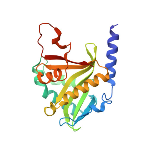
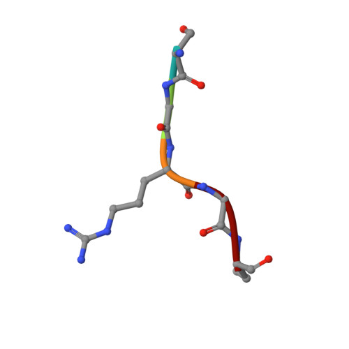
    Protein N-terminal acetyltransferase D (NatD, NAA40) that specifically acetylates the alpha-N-terminus of histone H4 and H2A has been implicated in various diseases, but no inhibitor has been reported for this important enzyme. Based on the acetyl transfer mechanism of NatD, we designed and prepared a series of highly potent NatD bisubstrate inhibitors by covalently linking coenzyme A to different peptide substrates via an acetyl or propionyl spacer. The most potent bisubstrate inhibitor displayed an apparent K i value of 1.0 nM. Biochemical studies indicated that bisubstrate inhibitors are competitive to the peptide substrate and noncompetitive to the cofactor, suggesting that NatD undergoes an ordered Bi-Bi mechanism. We also demonstrated that these inhibitors are highly specific toward NatD, displaying about 1000-fold selectivity over other closely related acetyltransferases. High-resolution crystal structures of NatD bound to two of these inhibitors revealed the molecular basis for their selectivity and inhibition mechanism, providing a rational path for future inhibitor development.


  • Organizational Affiliation
    • Department of Medicinal Chemistry and Molecular Pharmacology, Purdue Institute for Drug Discovery, Purdue University Center for Cancer Research, Purdue University, West Lafayette, Indiana 47907, United States.

Macromolecule Content 

  • Total Structure Weight: 51.05 kDa 
  • Atom Count: 3,820 
  • Modeled Residue Count: 416 
  • Deposited Residue Count: 418 
  • Unique protein chains: 2

Macromolecules

Find similar proteins by:|  3D Structure
Entity ID: 1
MoleculeChains  Sequence LengthOrganismDetailsImage
N-alpha-acetyltransferase 40A [auth D],
C [auth A]
204Homo sapiensMutation(s): 0 
Gene Names: NAA40NAT11PATT1
EC: 2.3.1.257
UniProt & NIH Common Fund Data Resources
Find proteins for Q86UY6 (Homo sapiens)
Explore Q86UY6 
Go to UniProtKB:  Q86UY6
PHAROS:  Q86UY6
GTEx:  ENSG00000110583 
Entity Groups
Sequence Clusters30% Identity50% Identity70% Identity90% Identity95% Identity100% Identity
UniProt GroupQ86UY6
Sequence Annotations
Expand
Reference Sequence
Find similar proteins by:  Sequence   |   3D Structure  
Entity ID: 2
MoleculeChains  Sequence LengthOrganismDetailsImage
SER-GLY-ARG-GLY-LYSB [auth E],
D [auth B]
5Homo sapiensMutation(s): 0 

Small Molecules

Ligands 5 Unique
IDChains Name / Formula / InChI Key2D Diagram3D Interactions
CMC
(Subject of Investigation/LOI)

Query on CMC


Download:Ideal Coordinates CCD File
K [auth E],
P [auth B]
CARBOXYMETHYL COENZYME *A
C23 H38 N7 O18 P3 S
OBUOSIHPWVNVJN-GRFIIANRSA-N
BTB

Query on BTB


Download:Ideal Coordinates CCD File
J [auth D]2-[BIS-(2-HYDROXY-ETHYL)-AMINO]-2-HYDROXYMETHYL-PROPANE-1,3-DIOL
C8 H19 N O5
OWMVSZAMULFTJU-UHFFFAOYSA-N
SO4

Query on SO4


Download:Ideal Coordinates CCD File
G [auth D],
H [auth D],
I [auth D],
N [auth A]
SULFATE ION
O4 S
QAOWNCQODCNURD-UHFFFAOYSA-L
GOL

Query on GOL


Download:Ideal Coordinates CCD File
E [auth D],
F [auth D],
M [auth A],
O [auth A]
GLYCEROL
C3 H8 O3
PEDCQBHIVMGVHV-UHFFFAOYSA-N
NH2

Query on NH2


Download:Ideal Coordinates CCD File
L [auth E],
Q [auth B]
AMINO GROUP
H2 N
QGZKDVFQNNGYKY-UHFFFAOYSA-N

Experimental Data & Validation

Experimental Data

  • Method: X-RAY DIFFRACTION
  • Resolution: 1.44 Å
  • R-Value Free:  0.183 (Depositor), 0.183 (DCC) 
  • R-Value Work:  0.161 (Depositor), 0.166 (DCC) 
  • R-Value Observed: 0.163 (Depositor) 
Space Group: P 21 21 21
Unit Cell:
Length ( Å )Angle ( ˚ )
a = 46.433α = 90
b = 74.349β = 90
c = 126.821γ = 90
Software Package:
Software NamePurpose
PHENIXrefinement
HKL-2000data reduction
HKL-2000data scaling
PHENIXphasing

Structure Validation

View Full Validation Report


Ligand Structure Quality Assessment 


Entry History 

& Funding Information

Deposition Data


Funding OrganizationLocationGrant Number
National Institutes of Health/National Institute of General Medical Sciences (NIH/NIGMS)United StatesR35 GM118090

Revision History  (Full details and data files)

  • Version 1.0: 2021-06-16
    Type: Initial release
  • Version 1.1: 2021-06-30
    Changes: Database references
  • Version 1.2: 2021-07-14
    Changes: Database references
  • Version 1.3: 2023-10-18
    Changes: Data collection, Database references, Refinement description