7KBU | pdb_00007kbu

Structure of Hevin FS-EC


Experimental Data Snapshot

  • Method: X-RAY DIFFRACTION
  • Resolution: 2.27 Å
  • R-Value Free: 
    0.238 (Depositor), 0.238 (DCC) 
  • R-Value Work: 
    0.186 (Depositor), 0.189 (DCC) 
  • R-Value Observed: 
    0.189 (Depositor) 

wwPDB Validation 3D Report Full Report

Validation slider image for 7KBU

This is version 1.3 of the entry. See complete history

Literature

Interplay between hevin, SPARC, and MDGAs: Modulators of neurexin-neuroligin transsynaptic bridges.

Fan, S.Gangwar, S.P.Machius, M.Rudenko, G.

(2021) Structure 29: 664

  • DOI: https://doi.org/10.1016/j.str.2021.01.003
  • Primary Citation Related Structures: 
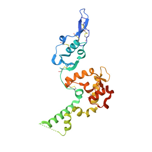
    7KBU

  • PubMed Abstract: 

    Hevin is secreted by astrocytes and its synaptogenic effects are antagonized by the related protein, SPARC. Hevin stabilizes neurexin-neuroligin transsynaptic bridges in vivo. A third protein, membrane-tethered MDGA, blocks these bridges. Here, we reveal the molecular underpinnings of a regulatory network formed by this trio of proteins. The hevin FS-EC structure differs from SPARC, in that the EC domain appears rearranged around a conserved core. The FS domain is structurally conserved and it houses nanomolar affinity binding sites for neurexin and neuroligin. SPARC also binds neurexin and neuroligin, competing with hevin, so its antagonist action is rooted in its shortened N-terminal region. Strikingly, the hevin FS domain competes with MDGA for an overlapping binding site on neuroligin, while the hevin EC domain binds the extracellular matrix protein collagen (like SPARC), so that this trio of proteins can regulate neurexin-neuroligin transsynaptic bridges and also extracellular matrix interactions, impacting synapse formation and ultimately neural circuits.


  • Organizational Affiliation
    • Department of Pharmacology and Toxicology, University of Texas Medical Branch, 301 University Boulevard, Galveston, TX 77555, USA; Sealy Center for Structural Biology and Molecular Biophysics, University of Texas Medical Branch, Galveston, TX 77555, USA.

Macromolecule Content 

  • Total Structure Weight: 58.55 kDa 
  • Atom Count: 3,632 
  • Modeled Residue Count: 413 
  • Deposited Residue Count: 488 
  • Unique protein chains: 1

Macromolecules

Find similar proteins by:|  3D Structure
Entity ID: 1
MoleculeChains  Sequence LengthOrganismDetailsImage
Proliferation-inducing protein 33
A, B
244Homo sapiensMutation(s): 0 
Gene Names: SPARCL1PIG33
UniProt & NIH Common Fund Data Resources
Find proteins for Q14515 (Homo sapiens)
Explore Q14515 
Go to UniProtKB:  Q14515
PHAROS:  Q14515
GTEx:  ENSG00000152583 
Entity Groups
Sequence Clusters30% Identity50% Identity70% Identity90% Identity95% Identity100% Identity
UniProt GroupQ14515
Glycosylation
Glycosylation Sites: 1Go to GlyGen: Q14515-1
Sequence Annotations
Expand
Reference Sequence

Oligosaccharides

Help  
Entity ID: 2
MoleculeChains Length2D Diagram GlycosylationD Interactions
2-acetamido-2-deoxy-beta-D-glucopyranose-(1-4)-2-acetamido-2-deoxy-beta-D-glucopyranose
C, D
2N-Glycosylation
Glycosylation Resources
GlyTouCan: G42666HT
GlyCosmos: G42666HT
GlyGen: G42666HT

Small Molecules

Ligands 4 Unique
IDChains Name / Formula / InChI Key2D Diagram3D Interactions
FMT

Query on FMT


Download:Ideal Coordinates CCD File
G [auth A],
O [auth B],
P [auth B],
Q [auth B]
FORMIC ACID
C H2 O2
BDAGIHXWWSANSR-UHFFFAOYSA-N
CA

Query on CA


Download:Ideal Coordinates CCD File
E [auth A]
F [auth A]
K [auth B]
L [auth B]
M [auth B]
E [auth A],
F [auth A],
K [auth B],
L [auth B],
M [auth B],
N [auth B]
CALCIUM ION
Ca
BHPQYMZQTOCNFJ-UHFFFAOYSA-N
CL

Query on CL


Download:Ideal Coordinates CCD File
H [auth A],
I [auth A],
J [auth A],
S [auth B]
CHLORIDE ION
Cl
VEXZGXHMUGYJMC-UHFFFAOYSA-M
NA

Query on NA


Download:Ideal Coordinates CCD File
R [auth B]SODIUM ION
Na
FKNQFGJONOIPTF-UHFFFAOYSA-N

Experimental Data & Validation

Experimental Data

  • Method: X-RAY DIFFRACTION
  • Resolution: 2.27 Å
  • R-Value Free:  0.238 (Depositor), 0.238 (DCC) 
  • R-Value Work:  0.186 (Depositor), 0.189 (DCC) 
  • R-Value Observed: 0.189 (Depositor) 
Space Group: I 21 21 21
Unit Cell:
Length ( Å )Angle ( ˚ )
a = 57.657α = 90
b = 132.289β = 90
c = 149.225γ = 90
Software Package:
Software NamePurpose
PHENIXrefinement
HKL-2000data reduction
HKL-2000data scaling
PDB_EXTRACTdata extraction
AutoSolphasing

Structure Validation

View Full Validation Report


Entry History 

& Funding Information

Deposition Data


Funding OrganizationLocationGrant Number
National Institutes of Health/National Institute of Mental Health (NIH/NIMH)United StatesR01MH077303

Revision History  (Full details and data files)

  • Version 1.0: 2021-02-10
    Type: Initial release
  • Version 1.1: 2021-02-17
    Changes: Database references
  • Version 1.2: 2021-07-14
    Changes: Database references
  • Version 1.3: 2024-11-06
    Changes: Data collection, Database references, Structure summary