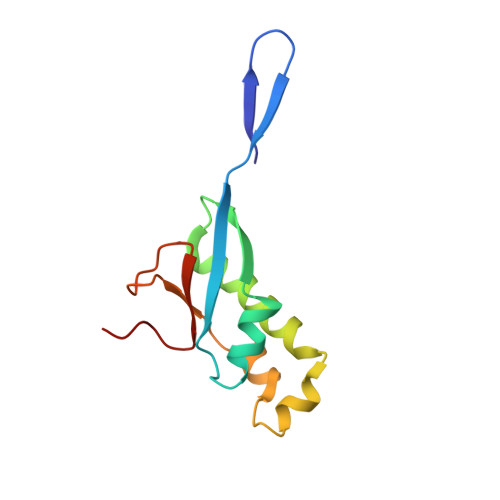
7KAG | pdb_00007kag

Crystal structure of the ubiquitin-like domain 1 (Ubl1) of Nsp3 from SARS-CoV-2


Experimental Data Snapshot

  • Method: X-RAY DIFFRACTION
  • Resolution: 3.21 Å
  • R-Value Free: 
    0.248 (Depositor), 0.252 (DCC) 
  • R-Value Work: 
    0.214 (Depositor), 0.216 (DCC) 
  • R-Value Observed: 
    0.217 (Depositor) 

Starting Model: experimental
View more details

wwPDB Validation 3D Report Full Report

Validation slider image for 7KAG

This is version 2.2 of the entry. See complete history

Literature

Structural and functional analyses of SARS-CoV-2 Nsp3 and its specific interactions with the 5' UTR of the viral genome.

Lemak, S.Skarina, T.Patel, D.T.Stogios, P.J.Savchenko, A.

(2025) Microbiol Spectr 13: e0287124-e0287124

  • DOI: https://doi.org/10.1128/spectrum.02871-24
  • Primary Citation Related Structures: 
    7KAG, 7LGO, 7RQG, 7T9W, 7TI9

  • PubMed Abstract: 

    Non-structural protein 3 (Nsp3) is the largest open reading frame encoded in the SARS-CoV-2 genome, essential for the formation of double-membrane vesicles (DMV) wherein viral RNA replication occurs. We conducted an extensive structure-function analysis of Nsp3 and determined the crystal structures of the ubiquitin-like 1 (Ubl1), nucleic acid binding (NAB), β-coronavirus-specific marker (βSM) domains, and a sub-region of the Y domain of this protein. We show that the Ubl1, ADP-ribose phosphatase (ADRP), human SARS Unique (HSUD), NAB, and Y domains of Nsp3 bind the 5' UTR of the viral genome and that the Ubl1 and Y domains possess affinity for recognition of this region, suggesting high specificity. The Ubl1-Nucleocapsid (N) protein complex binds the 5' UTR with greater affinity than the individual proteins alone. Our results suggest that multiple domains of Nsp3, particularly Ubl1 and Y, shepherd the 5' UTR of the viral genome during translocation through the DMV membrane, priming the Ubl1 domain to load the genome onto N protein.IMPORTANCEThe largest protein encoded by the SARS-CoV-2 genome is Nsp3. In infected cells, this multi-domain protein forms a pore structure in the virus-induced double-membrane vesicles (DMV). We have incomplete data on Nsp3 molecular structure, and here, we describe crystal structures for multiple domains of Nsp3. It is thought that newly replicated viral RNA transits through the DMV pore; however, we possess incomplete data on which regions of Nsp3 actually interact with RNA. Here, we present data showing that five domains of Nsp3 interact with the 5' UTR of the SARS-CoV-2 RNA, including the Y domain for which no function has ever been discovered. These data suggest that the pore structure plays an active role in recognizing the terminal end of the genome, transiting and loading the viral RNA onto the cytoplasmic nucleocapsid protein. These data help expand our knowledge of Nsp3 structure and function and the SARS-CoV-2 replication cycle.


  • Organizational Affiliation
    • BioZone, Department of Chemical Engineering and Applied Chemistry, University of Toronto, Toronto, Canada.

Macromolecule Content 

  • Total Structure Weight: 25.84 kDa 
  • Atom Count: 1,829 
  • Modeled Residue Count: 222 
  • Deposited Residue Count: 222 
  • Unique protein chains: 1

Macromolecules

Find similar proteins by:|  3D Structure
Entity ID: 1
MoleculeChains  Sequence LengthOrganismDetailsImage
Non-structural protein 3
A, B
111Severe acute respiratory syndrome coronavirus 2Mutation(s): 0 
Gene Names: rep1a-1b
EC: 3.4.22
UniProt
Find proteins for P0DTD1 (Severe acute respiratory syndrome coronavirus 2)
Explore P0DTD1 
Go to UniProtKB:  P0DTD1
Entity Groups
Sequence Clusters30% Identity50% Identity70% Identity90% Identity95% Identity100% Identity
UniProt GroupP0DTD1
Sequence Annotations
Expand
Reference Sequence

Small Molecules

Ligands 2 Unique
IDChains Name / Formula / InChI Key2D Diagram3D Interactions
SO4

Query on SO4


Download:Ideal Coordinates CCD File
L [auth B]SULFATE ION
O4 S
QAOWNCQODCNURD-UHFFFAOYSA-L
EDO

Query on EDO


Download:Ideal Coordinates CCD File
C [auth A]
D [auth A]
E [auth A]
F [auth A]
G [auth A]
C [auth A],
D [auth A],
E [auth A],
F [auth A],
G [auth A],
H [auth A],
I [auth B],
J [auth B],
K [auth B],
M [auth B],
N [auth B],
O [auth B],
P [auth B]
1,2-ETHANEDIOL
C2 H6 O2
LYCAIKOWRPUZTN-UHFFFAOYSA-N

Experimental Data & Validation

Experimental Data

  • Method: X-RAY DIFFRACTION
  • Resolution: 3.21 Å
  • R-Value Free:  0.248 (Depositor), 0.252 (DCC) 
  • R-Value Work:  0.214 (Depositor), 0.216 (DCC) 
  • R-Value Observed: 0.217 (Depositor) 
Space Group: C 1 2 1
Unit Cell:
Length ( Å )Angle ( ˚ )
a = 81.83α = 90
b = 147.57β = 125.896
c = 70.137γ = 90
Software Package:
Software NamePurpose
PHENIXrefinement
HKL-3000data reduction
HKL-3000data scaling
PHENIXphasing
PHENIXmodel building
Cootmodel building

Structure Validation

View Full Validation Report


Entry History 

& Funding Information

Deposition Data


Funding OrganizationLocationGrant Number
National Institutes of Health/National Institute Of Allergy and Infectious Diseases (NIH/NIAID)United StatesHHSN272201700060C

Revision History  (Full details and data files)

  • Version 1.0: 2020-10-14
    Type: Initial release
  • Version 1.1: 2021-01-27
    Changes: Structure summary
  • Version 2.0: 2022-01-26
    Changes: Database references, Polymer sequence
  • Version 2.1: 2023-10-18
    Changes: Data collection, Refinement description
  • Version 2.2: 2026-07-29
    Changes: Database references, Structure summary