Find PDB structures and Computed Structure Models (CSM) by combining queries from tools in this suite: Attribute Search, Sequence Similarity, Sequence Motif, 3D Similarity, and 3D Motif with 'AND' logic.
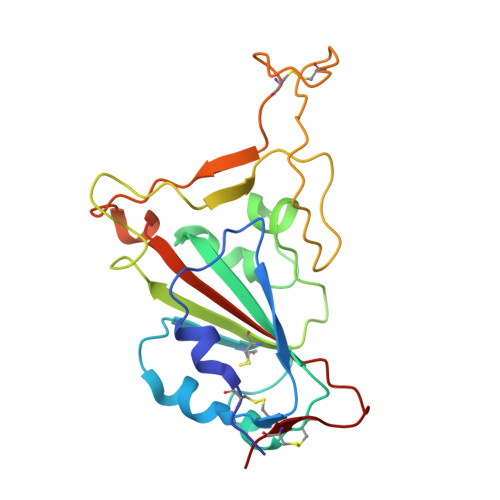
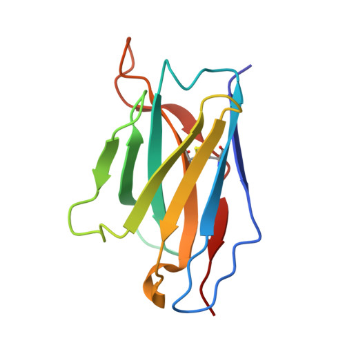
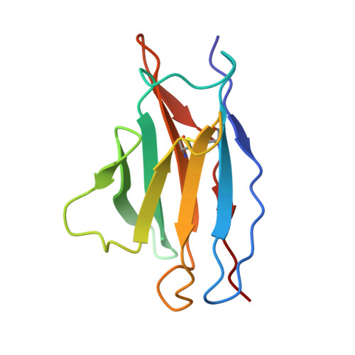
The severe acute respiratory syndrome coronavirus 2 (SARS-CoV-2) pandemic has necessitated the rapid development of antibody-based therapies and vaccines as countermeasures. Here, we use cryoelectron microscopy (cryo-EM) to characterize two protective anti-SARS-CoV-2 murine monoclonal antibodies (mAbs) in complex with the spike protein, revealing similarities between epitopes targeted by human and murine B cells. The more neutralizing mAb, 2B04, binds the receptor-binding motif (RBM) of the receptor-binding domain (RBD) and competes with angiotensin-converting enzyme 2 (ACE2). By contrast, 2H04 binds adjacent to the RBM and does not compete for ACE2 binding. Naturally occurring sequence variants of SARS-CoV-2 and corresponding neutralization escape variants selected in vitro map to our structurally defined epitopes, suggesting that SARS-CoV-2 might evade therapeutic antibodies with a limited set of mutations, underscoring the importance of combination mAb therapeutics. Finally, we show that 2B04 neutralizes SARS-CoV-2 infection by preventing ACE2 engagement, whereas 2H04 reduces host cell attachment without directly disrupting ACE2-RBM interactions, providing distinct inhibitory mechanisms used by RBD-specific mAbs.
Organizational Affiliation:
Department of Pathology & Immunology, Washington University School of Medicine, St. Louis, MO, USA.
Department of Pathology & Immunology, Washington University School of Medicine, St. Louis, MO, USA; Department of Medicine, Washington University School of Medicine, St. Louis, MO, USA.
Department of Molecular Microbiology, Washington University School of Medicine, St. Louis, MO, USA.
Department of Medicine, Washington University School of Medicine, St. Louis, MO, USA.
Washington University Center for Cellular Imaging, Washington University School of Medicine, St. Louis, MO, USA.
Washington University Center for Cellular Imaging, Washington University School of Medicine, St. Louis, MO, USA; Departments of Neuroscience and Cell Biology and Physiology, Washington University School of Medicine, St. Louis, MO, USA; Department of Biomedical Engineering, Washington University in St. Louis, St. Louis, MO, USA.
Department of Biochemistry and Molecular Biology, University of Texas Medical Branch, Galveston, TX, USA; Sealy Center for Structural Biology and Molecular Biophysics, University of Texas Medical Branch, Galveston, TX, USA; Sealy Institute for Vaccine Sciences, University of Texas Medical Branch, Galveston, TX, USA.
Department of Pathology & Immunology, Washington University School of Medicine, St. Louis, MO, USA; Department of Molecular Microbiology, Washington University School of Medicine, St. Louis, MO, USA; Department of Medicine, Washington University School of Medicine, St. Louis, MO, USA; The Andrew M. and Jane M. Bursky Center for Human Immunology & Immunotherapy Programs, Washington University School of Medicine, St. Louis, MO, USA.
Department of Pathology & Immunology, Washington University School of Medicine, St. Louis, MO, USA; Department of Molecular Microbiology, Washington University School of Medicine, St. Louis, MO, USA; The Andrew M. and Jane M. Bursky Center for Human Immunology & Immunotherapy Programs, Washington University School of Medicine, St. Louis, MO, USA.
Department of Pathology & Immunology, Washington University School of Medicine, St. Louis, MO, USA; Department of Molecular Microbiology, Washington University School of Medicine, St. Louis, MO, USA; The Andrew M. and Jane M. Bursky Center for Human Immunology & Immunotherapy Programs, Washington University School of Medicine, St. Louis, MO, USA; Department of Biochemistry & Molecular Biophysics, Washington University School of Medicine, St. Louis, MO, USA. Electronic address: fremont@wustl.edu.

 Download Mendeley