Cryo-EM structure of the EspA filament from enteropathogenic Escherichia coli: Revealing the mechanism of effector translocation in the T3SS.
Lyons, B.J.E., Atkinson, C.E., Deng, W., Serapio-Palacios, A., Finlay, B.B., Strynadka, N.C.J.(2021) Structure 29: 479
- PubMed: 33453150 Search on PubMed
- DOI: https://doi.org/10.1016/j.str.2020.12.009
- Primary Citation Related Structures:
7K7K - PubMed Abstract:
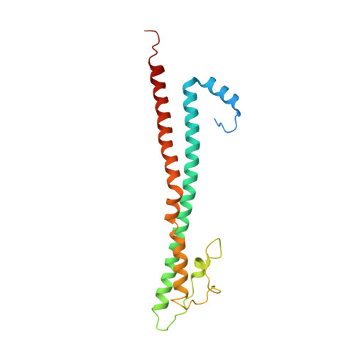
The type III secretion system (T3SS) is a virulence mechanism employed by Gram-negative pathogens. The T3SS forms a proteinaceous channel that projects a needle into the extracellular medium where it interacts with the host cell to deliver virulence factors. Enteropathogenic Escherichia coli (EPEC) is unique in adopting a needle extension to the T3SS-a filament formed by EspA-which is absolutely required for efficient colonization of the gut. Here, we describe the cryoelectron microscopy structure of native EspA filaments from EPEC at 3.6-Å resolution. Within the filament, positively charged residues adjacent to a hydrophobic groove line the lumen of the filament in a spiral manner, suggesting a mechanism of substrate translocation mediated via electrostatics. Using structure-guided mutagenesis, in vivo studies corroborate the role of these residues in secretion and translocation function. The high-resolution structure of the EspA filament could aid in structure-guided drug design of antivirulence therapeutics.
- Department of Biochemistry and Molecular Biology, University of British Columbia, Vancouver, BC V6T 1Z3, Canada; Centre for Blood Research, University of British Columbia, Vancouver, BC V6T 1Z3, Canada.
Organizational Affiliation: