7K57 | pdb_00007k57

Structure of apo VCP dodecamer generated from bacterially recombinant VCP/p97


Experimental Data Snapshot

  • Method: ELECTRON MICROSCOPY
  • Resolution: 3.70 Å
  • Aggregation State: PARTICLE 
  • Reconstruction Method: SINGLE PARTICLE 

wwPDB Validation 3D Report Full Report

Validation slider image for 7K57

This is version 1.2 of the entry. See complete history

Literature

Cryo-electron microscopy structures of VCP/p97 reveal a new mechanism of oligomerization regulation.

Yu, G.Bai, Y.Li, K.Amarasinghe, O.Jiang, W.Zhang, Z.Y.

(2021) iScience 24: 103310-103310

  • DOI: https://doi.org/10.1016/j.isci.2021.103310
  • Primary Citation Related Structures: 
    7K56, 7K57, 7K59

  • PubMed Abstract: 

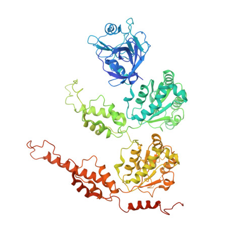
    VCP/p97 is an evolutionarily conserved AAA+ ATPase important for cellular homeostasis. Previous studies suggest that VCP predominantly exists as a homohexamer. Here, we performed structural and biochemical characterization of VCP dodecamer, an understudied state of VCP. The structure revealed an apo nucleotide status that has rarely been captured, a tail-to-tail assembly of two hexamers, and the up-elevated N-terminal domains akin to that seen in the ATP-bound hexamer. Further analyses elucidated a nucleotide status-dependent dodecamerization mechanism, where nucleotide dissociation from the D2 AAA domains induces and promotes VCP dodecamerization. In contrast, nucleotide-free D1 AAA domains are associated with the up-rotation of N-terminal domains, which may prime D1 for ATP binding. These results therefore reveal new nucleotide status-dictated intra- and interhexamer conformational changes and suggest that modulation of D2 domain nucleotide occupancy may serve as a mechanism in controlling VCP oligomeric states.


  • Organizational Affiliation
    • Departments of Medicinal Chemistry and Molecular Pharmacology and of Chemistry, Center for Cancer Research, and Institute for Drug Discovery, Purdue University, 720 Clinic Drive, West Lafayette, IN 47907, USA.

Macromolecule Content 

  • Total Structure Weight: 1,073.58 kDa 
  • Atom Count: 70,104 
  • Modeled Residue Count: 8,892 
  • Deposited Residue Count: 9,672 
  • Unique protein chains: 1

Macromolecules

Find similar proteins by:|  3D Structure
Entity ID: 1
MoleculeChains  Sequence LengthOrganismDetailsImage
Transitional endoplasmic reticulum ATPase
A, B, C, D, E
A, B, C, D, E, F, G, H, I, J, K, L
806Homo sapiensMutation(s): 0 
Gene Names: VCP
EC: 3.6.4.6
UniProt & NIH Common Fund Data Resources
Find proteins for P55072 (Homo sapiens)
Explore P55072 
Go to UniProtKB:  P55072
PHAROS:  P55072
GTEx:  ENSG00000165280 
Entity Groups
Sequence Clusters30% Identity50% Identity70% Identity90% Identity95% Identity100% Identity
UniProt GroupP55072
Sequence Annotations
Expand
Reference Sequence

Experimental Data & Validation

Experimental Data

  • Method: ELECTRON MICROSCOPY
  • Resolution: 3.70 Å
  • Aggregation State: PARTICLE 
  • Reconstruction Method: SINGLE PARTICLE 
EM Software:
TaskSoftware PackageVersion
RECONSTRUCTIONcryoSPARC

Structure Validation

View Full Validation Report


Entry History 

& Funding Information

Deposition Data


Funding OrganizationLocationGrant Number
National Institutes of Health/National Cancer Institute (NIH/NCI)United StatesRO1 CA69202

Revision History  (Full details and data files)

  • Version 1.0: 2021-10-13
    Type: Initial release
  • Version 1.1: 2022-04-06
    Changes: Database references
  • Version 1.2: 2024-05-29
    Changes: Data collection