7K3M | pdb_00007k3m

Crystal Structure of the Beta Lactamase Class D from Chitinophaga pinensis by Serial Crystallography


Experimental Data Snapshot

  • Method: X-RAY DIFFRACTION
  • Resolution: 1.80 Å
  • R-Value Free: 
    0.224 (Depositor), 0.226 (DCC) 
  • R-Value Work: 
    0.177 (Depositor), 0.177 (DCC) 
  • R-Value Observed: 
    0.183 (Depositor) 

Starting Model: experimental
View more details

wwPDB Validation 3D Report Full Report

Validation slider image for 7K3M

This is version 1.4 of the entry. See complete history

Literature

Fixed-target serial crystallography at the Structural Biology Center.

Sherrell, D.A.Lavens, A.Wilamowski, M.Kim, Y.Chard, R.Lazarski, K.Rosenbaum, G.Vescovi, R.Johnson, J.L.Akins, C.Chang, C.Michalska, K.Babnigg, G.Foster, I.Joachimiak, A.

(2022) J Synchrotron Radiat 29: 1141-1151

  • DOI: https://doi.org/10.1107/S1600577522007895
  • Primary Citation Related Structures: 
    7K3M, 7L52

  • PubMed Abstract: 

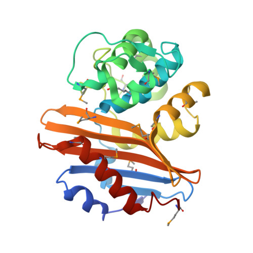
    Serial synchrotron crystallography enables the study of protein structures under physiological temperature and reduced radiation damage by collection of data from thousands of crystals. The Structural Biology Center at Sector 19 of the Advanced Photon Source has implemented a fixed-target approach with a new 3D-printed mesh-holder optimized for sample handling. The holder immobilizes a crystal suspension or droplet emulsion on a nylon mesh, trapping and sealing a near-monolayer of crystals in its mother liquor between two thin Mylar films. Data can be rapidly collected in scan mode and analyzed in near real-time using piezoelectric linear stages assembled in an XYZ arrangement, controlled with a graphical user interface and analyzed using a high-performance computing pipeline. Here, the system was applied to two β-lactamases: a class D serine β-lactamase from Chitinophaga pinensis DSM 2588 and L1 metallo-β-lactamase from Stenotrophomonas maltophilia K279a.


  • Organizational Affiliation
    • Structural Biology Center, X-ray Science Division, Argonne National Laboratory, Lemont, IL 60439, USA.

Macromolecule Content 

  • Total Structure Weight: 29.88 kDa 
  • Atom Count: 2,160 
  • Modeled Residue Count: 245 
  • Deposited Residue Count: 250 
  • Unique protein chains: 1

Macromolecules

Find similar proteins by:|  3D Structure
Entity ID: 1
MoleculeChains  Sequence LengthOrganismDetailsImage
Beta-lactamase250Chitinophaga pinensis DSM 2588Mutation(s): 0 
Gene Names: Cpin_0907
EC: 3.5.2.6

Small Molecules

Modified Residues  2 Unique
IDChains TypeFormula2D DiagramParent
KCX
Query on KCX
A
L-PEPTIDE LINKINGC7 H14 N2 O4LYS
MSE
Query on MSE
A
L-PEPTIDE LINKINGC5 H11 N O2 SeMET

Experimental Data & Validation

Experimental Data

  • Method: X-RAY DIFFRACTION
  • Resolution: 1.80 Å
  • R-Value Free:  0.224 (Depositor), 0.226 (DCC) 
  • R-Value Work:  0.177 (Depositor), 0.177 (DCC) 
  • R-Value Observed: 0.183 (Depositor) 
Space Group: P 21 21 21
Unit Cell:
Length ( Å )Angle ( ˚ )
a = 49.413α = 90
b = 69.347β = 90
c = 70.035γ = 90
Software Package:
Software NamePurpose
PHENIXrefinement
CRISpydata collection
DIALSdata reduction
PRIMEdata scaling
MOLREPphasing

Structure Validation

View Full Validation Report


Entry History 

& Funding Information

Deposition Data


Funding OrganizationLocationGrant Number
National Institutes of Health/National Institute Of Allergy and Infectious Diseases (NIH/NIAID)United States--

Revision History  (Full details and data files)

  • Version 1.0: 2020-09-23
    Type: Initial release
  • Version 1.1: 2020-10-28
    Changes: Refinement description
  • Version 1.2: 2020-12-23
    Changes: Database references, Structure summary
  • Version 1.3: 2024-04-03
    Changes: Data collection, Database references, Refinement description
  • Version 1.4: 2026-03-25
    Changes: Database references, Structure summary