7K26 | pdb_00007k26

Crystal structure of Human H-chain Ferritin variant infused with Sodium Acrylate


Experimental Data Snapshot

  • Method: X-RAY DIFFRACTION
  • Resolution: 2.70 Å
  • R-Value Free: 
    0.254 (Depositor), 0.252 (DCC) 
  • R-Value Work: 
    0.198 (Depositor), 0.199 (DCC) 
  • R-Value Observed: 
    0.201 (Depositor) 

Starting Model: experimental
View more details

wwPDB Validation 3D Report Full Report

Validation slider image for 7K26

This is version 1.2 of the entry. See complete history

Literature

Anisotropic Dynamics and Mechanics of Macromolecular Crystals Containing Lattice-Patterned Polymer Networks.

Han, K.Bailey, J.B.Zhang, L.Tezcan, F.A.

(2020) J Am Chem Soc 142: 19402-19410

  • DOI: https://doi.org/10.1021/jacs.0c10065
  • Primary Citation Related Structures: 
    6WYF, 6WYG, 6WYH, 7K26

  • PubMed Abstract: 

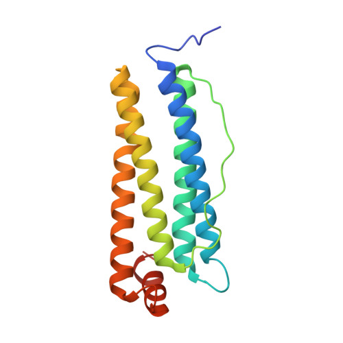
    The mechanical and functional properties of many crystalline materials depend on cooperative changes in lattice arrangements in response to external perturbations. However, the flexibility and adaptiveness of crystalline materials are limited. Additionally, the bottom-up, molecular-level design of crystals with desired dynamic and mechanical properties at the macroscopic level remains a considerable challenge. To address these challenges, we had previously integrated mesoporous, cubic ferritin crystals with hydrogel networks, resulting in hybrid materials (polymer-integrated crystals or PIX) which could undergo dramatic structural changes while maintaining crystalline periodicity and display efficient self-healing. The dynamics and mechanics of these ferritin-PIX were devoid of directionality, which is an important attribute of many molecular and macroscopic materials/devices. In this study, we report that such directionality can be achieved through the use of ferritin crystals with anisotropic symmetries (rhombohedral or trigonal), which enable the templated formation of patterned hydrogel networks in crystallo. The resulting PIX expand and contract anisotropically without losing crystallinity, undergo prompt bending motions in response to stimuli, and self-heal efficiently, capturing some of the essential features of sophisticated biological devices like skeletal muscles.


  • Organizational Affiliation
    • Department of Chemistry and Biochemistry, University of California, San Diego, La Jolla, California 92093, United States.

Macromolecule Content 

  • Total Structure Weight: 254.68 kDa 
  • Atom Count: 17,300 
  • Modeled Residue Count: 2,084 
  • Deposited Residue Count: 2,184 
  • Unique protein chains: 1

Macromolecules

Find similar proteins by:|  3D Structure
Entity ID: 1
MoleculeChains  Sequence LengthOrganismDetailsImage
Ferritin heavy chain
A, B, C, D, E
A, B, C, D, E, F, G, H, I, J, K, L
182Homo sapiensMutation(s): 0 
Gene Names: FTH1FTHFTHL6OK/SW-cl.84PIG15
EC: 1.16.3.1
UniProt & NIH Common Fund Data Resources
Find proteins for P02794 (Homo sapiens)
Explore P02794 
Go to UniProtKB:  P02794
PHAROS:  P02794
GTEx:  ENSG00000167996 
Entity Groups
Sequence Clusters30% Identity50% Identity70% Identity90% Identity95% Identity100% Identity
UniProt GroupP02794
Sequence Annotations
Expand
Reference Sequence

Small Molecules

Ligands 3 Unique
IDChains Name / Formula / InChI Key2D Diagram3D Interactions
MPD

Query on MPD


Download:Ideal Coordinates CCD File
AA [auth F]
CA [auth G]
IA [auth K]
LA [auth L]
S [auth B]
AA [auth F],
CA [auth G],
IA [auth K],
LA [auth L],
S [auth B],
W [auth D],
Y [auth E]
(4S)-2-METHYL-2,4-PENTANEDIOL
C6 H14 O2
SVTBMSDMJJWYQN-YFKPBYRVSA-N
FE

Query on FE


Download:Ideal Coordinates CCD File
BA [auth G]
DA [auth H]
FA [auth I]
GA [auth J]
HA [auth K]
BA [auth G],
DA [auth H],
FA [auth I],
GA [auth J],
HA [auth K],
JA [auth L],
M [auth A],
P [auth B],
T [auth C],
V [auth D],
X [auth E],
Z [auth F]
FE (III) ION
Fe
VTLYFUHAOXGGBS-UHFFFAOYSA-N
NA

Query on NA


Download:Ideal Coordinates CCD File
EA [auth H]
KA [auth L]
N [auth A]
O [auth A]
Q [auth B]
EA [auth H],
KA [auth L],
N [auth A],
O [auth A],
Q [auth B],
R [auth B],
U [auth C]
SODIUM ION
Na
FKNQFGJONOIPTF-UHFFFAOYSA-N

Experimental Data & Validation

Experimental Data

  • Method: X-RAY DIFFRACTION
  • Resolution: 2.70 Å
  • R-Value Free:  0.254 (Depositor), 0.252 (DCC) 
  • R-Value Work:  0.198 (Depositor), 0.199 (DCC) 
  • R-Value Observed: 0.201 (Depositor) 
Space Group: P 31 2 1
Unit Cell:
Length ( Å )Angle ( ˚ )
a = 131.57α = 90
b = 131.57β = 90
c = 306.56γ = 120
Software Package:
Software NamePurpose
PHENIXrefinement
iMOSFLMdata reduction
Aimlessdata scaling
PHASERphasing

Structure Validation

View Full Validation Report


Entry History 

Deposition Data

Revision History  (Full details and data files)

  • Version 1.0: 2020-11-11
    Type: Initial release
  • Version 1.1: 2020-11-25
    Changes: Database references
  • Version 1.2: 2023-10-18
    Changes: Data collection, Database references, Refinement description