7K0Z | pdb_00007k0z

Marsupial T cell receptor Spl_157


Experimental Data Snapshot

  • Method: X-RAY DIFFRACTION
  • Resolution: 3.20 Å
  • R-Value Free: 
    0.285 (Depositor), 0.300 (DCC) 
  • R-Value Work: 
    0.257 (Depositor), 0.260 (DCC) 
  • R-Value Observed: 
    0.259 (Depositor) 

Starting Model: other
View more details

wwPDB Validation 3D Report Full Report

Validation slider image for 7K0Z

Ligand Structure Quality Assessment 


This is version 1.3 of the entry. See complete history

Literature

The molecular assembly of the marsupial gamma mu T cell receptor defines a third T cell lineage.

Morrissey, K.A.Wegrecki, M.Praveena, T.Hansen, V.L.Bu, L.Sivaraman, K.K.Darko, S.Douek, D.C.Rossjohn, J.Miller, R.D.Le Nours, J.

(2021) Science 371: 1383-1388

  • DOI: https://doi.org/10.1126/science.abe7070
  • Primary Citation Related Structures: 
    7K0X, 7K0Z, 7L15

  • PubMed Abstract: 

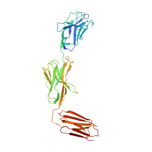
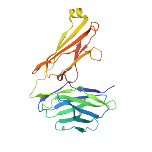
    αβ and γδ T cell receptors (TCRs) are highly diverse antigen receptors that define two evolutionarily conserved T cell lineages. We describe a population of γμTCRs found exclusively in non-eutherian mammals that consist of a two-domain (Vγ-Cγ) γ-chain paired to a three-domain (Vμ-Vμj-Cμ) μ-chain. γμTCRs were characterized by restricted diversity in the Vγ and Vμj domains and a highly diverse unpaired Vμ domain. Crystal structures of two distinct γμTCRs revealed the structural basis of the association of the γμTCR heterodimer. The Vμ domain shared the characteristics of a single-domain antibody within which the hypervariable CDR3μ loop suggests a major antigen recognition determinant. We define here the molecular basis underpinning the assembly of a third TCR lineage, the γμTCR.


  • Organizational Affiliation
    • Department of Biology, Center for Evolutionary and Theoretical Immunology, University of New Mexico, Albuquerque, NM, USA.

Macromolecule Content 

  • Total Structure Weight: 130.64 kDa 
  • Atom Count: 7,936 
  • Modeled Residue Count: 1,085 
  • Deposited Residue Count: 1,170 
  • Unique protein chains: 2

Macromolecules

Find similar proteins by:|  3D Structure
Entity ID: 1
MoleculeChains  Sequence LengthOrganismDetailsImage
T cell receptor mu chainA [auth C],
C [auth A]
349Monodelphis domesticaMutation(s): 0 
Find similar proteins by:|  3D Structure
Entity ID: 2
MoleculeChains  Sequence LengthOrganismDetailsImage
T cell receptor gamma chainB [auth D],
D [auth B]
236Monodelphis domesticaMutation(s): 0 

Small Molecules

Ligands 1 Unique
IDChains Name / Formula / InChI Key2D Diagram3D Interactions
NAG

Query on NAG


Download:Ideal Coordinates CCD File
E [auth C]2-acetamido-2-deoxy-beta-D-glucopyranose
C8 H15 N O6
OVRNDRQMDRJTHS-FMDGEEDCSA-N

Experimental Data & Validation

Experimental Data

  • Method: X-RAY DIFFRACTION
  • Resolution: 3.20 Å
  • R-Value Free:  0.285 (Depositor), 0.300 (DCC) 
  • R-Value Work:  0.257 (Depositor), 0.260 (DCC) 
  • R-Value Observed: 0.259 (Depositor) 
Space Group: P 21 21 21
Unit Cell:
Length ( Å )Angle ( ˚ )
a = 72.328α = 90
b = 110.722β = 90
c = 207.798γ = 90
Software Package:
Software NamePurpose
PHENIXrefinement
XDSdata reduction
Aimlessdata scaling
PHASERphasing

Structure Validation

View Full Validation Report


Ligand Structure Quality Assessment 


Entry History 

Deposition Data

Revision History  (Full details and data files)

  • Version 1.0: 2021-03-24
    Type: Initial release
  • Version 1.1: 2021-04-14
    Changes: Database references
  • Version 1.2: 2024-04-03
    Changes: Data collection, Database references, Refinement description
  • Version 1.3: 2024-10-30
    Changes: Structure summary