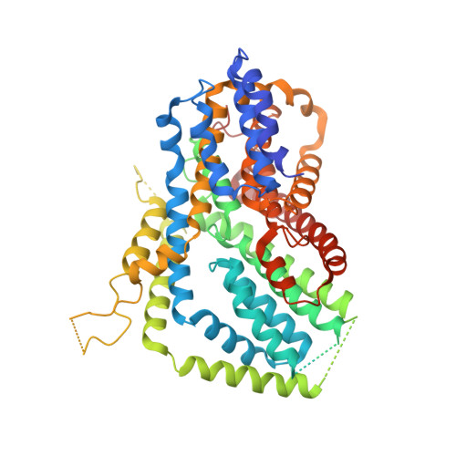
7JSK

Structure of the NaCT-Citrate complex

  • Classification: MEMBRANE PROTEIN
  • Organism(s): Homo sapiens
  • Expression System: Trichoplusia ni
  • Mutation(s): No 
  • Membrane Protein: Yes  OPMPDBTMMemProtMDmpstruc

  • Deposited: 2020-08-14 Released: 2021-02-24 
  • Deposition Author(s): Sauer, D.B., Wang, B., Song, J., Rice, W.J., Wang, D.N.
  • Funding Organization(s): National Institutes of Health/National Institute of Neurological Disorders and Stroke (NIH/NINDS), National Institutes of Health/National Institute of General Medical Sciences (NIH/NIGMS), American Cancer Society, Department of Defense (DOD, United States), The G. Harold and Leila Y. Mathers Foundation, TESS Research Foundation, American Epilepsy Society

Experimental Data Snapshot

  • Method: ELECTRON MICROSCOPY
  • Resolution: 3.04 Å
  • Aggregation State: PARTICLE 
  • Reconstruction Method: SINGLE PARTICLE 

wwPDB Validation   3D Report Full Report


This is version 1.3 of the entry. See complete history


Literature

Structure and inhibition mechanism of the human citrate transporter NaCT.

Sauer, D.B.Song, J.Wang, B.Hilton, J.K.Karpowich, N.K.Mindell, J.A.Rice, W.J.Wang, D.N.

(2021) Nature 591: 157-161

  • DOI: https://doi.org/10.1038/s41586-021-03230-x
  • Primary Citation of Related Structures:  
    7JSJ, 7JSK

  • PubMed Abstract: 

    Citrate is best known as an intermediate in the tricarboxylic acid cycle of the cell. In addition to this essential role in energy metabolism, the tricarboxylate anion also acts as both a precursor and a regulator of fatty acid synthesis 1-3 . Thus, the rate of fatty acid synthesis correlates directly with the cytosolic concentration of citrate 4,5 . Liver cells import citrate through the sodium-dependent citrate transporter NaCT (encoded by SLC13A5) and, as a consequence, this protein is a potential target for anti-obesity drugs. Here, to understand the structural basis of its inhibition mechanism, we determined cryo-electron microscopy structures of human NaCT in complexes with citrate or a small-molecule inhibitor. These structures reveal how the inhibitor-which binds to the same site as citrate-arrests the transport cycle of NaCT. The NaCT-inhibitor structure also explains why the compound selectively inhibits NaCT over two homologous human dicarboxylate transporters, and suggests ways to further improve the affinity and selectivity. Finally, the NaCT structures provide a framework for understanding how various mutations abolish the transport activity of NaCT in the brain and thereby cause epilepsy associated with mutations in SLC13A5 in newborns (which is known as SLC13A5-epilepsy) 6-8 .


  • Organizational Affiliation

    Skirball Institute of Biomolecular Medicine, New York University School of Medicine, New York, NY, USA.


Macromolecules
Find similar proteins by:  (by identity cutoff)  |  3D Structure
Entity ID: 1
MoleculeChains Sequence LengthOrganismDetailsImage
Solute carrier family 13 member 5
A, B
568Homo sapiensMutation(s): 0 
Gene Names: SLC13A5NACT
Membrane Entity: Yes 
UniProt & NIH Common Fund Data Resources
Find proteins for Q86YT5 (Homo sapiens)
Explore Q86YT5 
Go to UniProtKB:  Q86YT5
PHAROS:  Q86YT5
GTEx:  ENSG00000141485 
Entity Groups  
Sequence Clusters30% Identity50% Identity70% Identity90% Identity95% Identity100% Identity
UniProt GroupQ86YT5
Glycosylation
Glycosylation Sites: 1Go to GlyGen: Q86YT5-1
Sequence Annotations
Expand
  • Reference Sequence
Small Molecules
Ligands 3 Unique
IDChains Name / Formula / InChI Key2D Diagram3D Interactions
NAG
Query on NAG

Download Ideal Coordinates CCD File 
C [auth A],
G [auth B]
2-acetamido-2-deoxy-beta-D-glucopyranose
C8 H15 N O6
OVRNDRQMDRJTHS-FMDGEEDCSA-N
CIT (Subject of Investigation/LOI)
Query on CIT

Download Ideal Coordinates CCD File 
D [auth A],
H [auth B]
CITRIC ACID
C6 H8 O7
KRKNYBCHXYNGOX-UHFFFAOYSA-N
NA
Query on NA

Download Ideal Coordinates CCD File 
E [auth A],
F [auth A],
I [auth B],
J [auth B]
SODIUM ION
Na
FKNQFGJONOIPTF-UHFFFAOYSA-N
Experimental Data & Validation

Experimental Data

  • Method: ELECTRON MICROSCOPY
  • Resolution: 3.04 Å
  • Aggregation State: PARTICLE 
  • Reconstruction Method: SINGLE PARTICLE 
EM Software:
TaskSoftware PackageVersion
RECONSTRUCTIONcryoSPARC
MODEL REFINEMENTPHENIX

Structure Validation

View Full Validation Report


Entry History & Funding Information

Deposition Data


Funding OrganizationLocationGrant Number
National Institutes of Health/National Institute of Neurological Disorders and Stroke (NIH/NINDS)United StatesR01NS108151
National Institutes of Health/National Institute of General Medical Sciences (NIH/NIGMS)United StatesR01GM121994
American Cancer SocietyUnited States129844-PF-17-135-01-TBE
Department of Defense (DOD, United States)United StatesW81XWH-16-1-0153
The G. Harold and Leila Y. Mathers FoundationUnited States--
TESS Research FoundationUnited States--
American Epilepsy SocietyUnited States--

Revision History  (Full details and data files)

  • Version 1.0: 2021-02-24
    Type: Initial release
  • Version 1.1: 2021-03-03
    Changes: Data processing, Database references
  • Version 1.2: 2021-03-10
    Changes: Database references
  • Version 1.3: 2024-10-23
    Changes: Data collection, Database references, Structure summary