7JOC

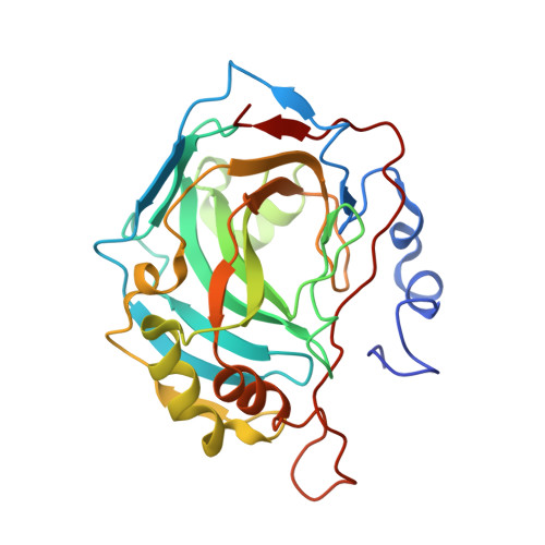
Carbonic Anhydrase IX Mimic Complexed with N-(5-sulfamoyl-1,3,4-thiadiazol-2-yl)adamantanecarboxamide


Experimental Data Snapshot

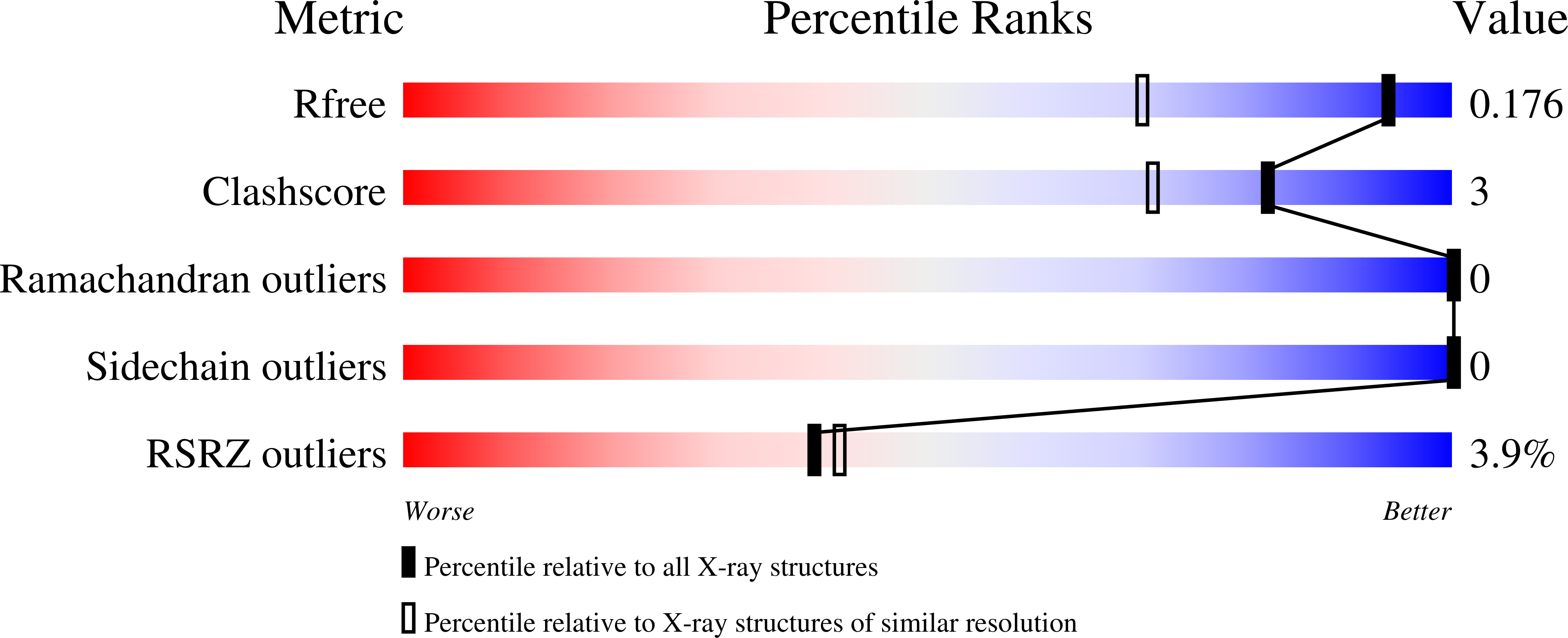
  • Method: X-RAY DIFFRACTION
  • Resolution: 1.39 Å
  • R-Value Free: 0.176 
  • R-Value Work: 0.157 
  • R-Value Observed: 0.158 

Starting Model: experimental
View more details

wwPDB Validation   3D Report Full Report


Ligand Structure Quality Assessment 


This is version 1.2 of the entry. See complete history


Literature

Structural Basis of Nanomolar Inhibition of Tumor-Associated Carbonic Anhydrase IX: X-Ray Crystallographic and Inhibition Study of Lipophilic Inhibitors with Acetazolamide Backbone.

Andring, J.T.Fouch, M.Akocak, S.Angeli, A.Supuran, C.T.Ilies, M.A.McKenna, R.

(2020) J Med Chem 63: 13064-13075

  • DOI: https://doi.org/10.1021/acs.jmedchem.0c01390
  • Primary Citation of Related Structures:  
    7JNR, 7JNV, 7JNW, 7JNX, 7JNZ, 7JO0, 7JO1, 7JO2, 7JO3, 7JOB, 7JOC

  • PubMed Abstract: 

    This study provides a structure-activity relationship study of a series of lipophilic carbonic anhydrase (CA) inhibitors with an acetazolamide backbone. The inhibitors were tested against the tumor-expressed CA isozyme IX (CA IX), and the cytosolic CA I, CA II, and membrane-bound CA IV. The study identified several low nanomolar potent inhibitors against CA IX, with lipophilicities spanning two log units. Very potent pan-inhibitors with nanomolar potency against CA IX and sub-nanomolar potency against CA II and CA IV, and with potency against CA I one order of magnitude better than the parent acetazolamide 1 were also identified in this study, together with compounds that displayed selectivity against membrane-bound CA IV. A comprehensive X-ray crystallographic study (12 crystal structures), involving both CA II and a soluble CA IX mimetic (CA IX-mimic), revealed the structural basis of this particular inhibition profile and laid the foundation for further developments toward more potent and selective inhibitors for the tumor-expressed CA IX.


  • Organizational Affiliation

    Department of Biochemistry and Molecular Biology, College of Medicine, University of Florida, Gainesville, Florida 32610, United States.


Macromolecules
Find similar proteins by:  (by identity cutoff)  |  3D Structure
Entity ID: 1
MoleculeChains Sequence LengthOrganismDetailsImage
Carbonic anhydrase 2260Homo sapiensMutation(s): 7 
Gene Names: CA2
EC: 4.2.1.1 (PDB Primary Data), 4.2.1.69 (UniProt)
UniProt & NIH Common Fund Data Resources
Find proteins for P00918 (Homo sapiens)
Explore P00918 
Go to UniProtKB:  P00918
PHAROS:  P00918
GTEx:  ENSG00000104267 
Entity Groups  
Sequence Clusters30% Identity50% Identity70% Identity90% Identity95% Identity100% Identity
UniProt GroupP00918
Sequence Annotations
Expand
  • Reference Sequence
Experimental Data & Validation

Experimental Data

  • Method: X-RAY DIFFRACTION
  • Resolution: 1.39 Å
  • R-Value Free: 0.176 
  • R-Value Work: 0.157 
  • R-Value Observed: 0.158 
  • Space Group: P 1 21 1
Unit Cell:
Length ( Å )Angle ( ˚ )
a = 42.134α = 90
b = 41.697β = 103.84
c = 72.375γ = 90
Software Package:
Software NamePurpose
PHENIXrefinement
Aimlessdata scaling
PDB_EXTRACTdata extraction
XDSdata reduction
PHENIXphasing

Structure Validation

View Full Validation Report


Ligand Structure Quality Assessment 


Entry History 

Deposition Data

Revision History  (Full details and data files)

  • Version 1.0: 2020-11-04
    Type: Initial release
  • Version 1.1: 2020-11-18
    Changes: Database references
  • Version 1.2: 2023-10-18
    Changes: Data collection, Database references, Refinement description