7JN3 | pdb_00007jn3

Cryo-EM structure of Rous sarcoma virus cleaved synaptic complex (CSC) with HIV-1 integrase strand transfer inhibitor MK-2048


Experimental Data Snapshot

  • Method: ELECTRON MICROSCOPY
  • Resolution: 3.21 Å
  • Aggregation State: PARTICLE 
  • Reconstruction Method: SINGLE PARTICLE 

Starting Model: experimental
View more details

wwPDB Validation 3D Report Full Report

Validation slider image for 7JN3

This is version 1.2 of the entry. See complete history

Literature

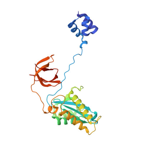
Cryo-EM structure of the Rous sarcoma virus octameric cleaved synaptic complex intasome.

Pandey, K.K.Bera, S.Shi, K.Rau, M.J.Oleru, A.V.Fitzpatrick, J.A.J.Engelman, A.N.Aihara, H.Grandgenett, D.P.

(2021) Commun Biol 4: 330-330

  • DOI: https://doi.org/10.1038/s42003-021-01855-2
  • Primary Citation Related Structures: 
    7JN3, 7KU7, 7KUI

  • PubMed Abstract: 

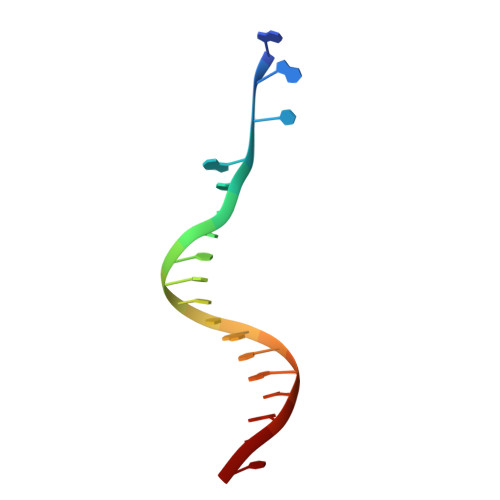
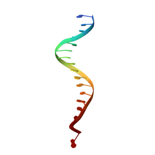
    Despite conserved catalytic integration mechanisms, retroviral intasomes composed of integrase (IN) and viral DNA possess diverse structures with variable numbers of IN subunits. To investigate intasome assembly mechanisms, we employed the Rous sarcoma virus (RSV) IN dimer that assembles a precursor tetrameric structure in transit to the mature octameric intasome. We determined the structure of RSV octameric intasome stabilized by a HIV-1 IN strand transfer inhibitor using single particle cryo-electron microscopy. The structure revealed significant flexibility of the two non-catalytic distal IN dimers along with previously unrecognized movement of the conserved intasome core, suggesting ordered conformational transitions between intermediates that may be important to capture the target DNA. Single amino acid substitutions within the IN C-terminal domain affected intasome assembly and function in vitro and infectivity of pseudotyped RSV virions. Unexpectedly, 17 C-terminal amino acids of IN were dispensable for virus infection despite regulating the transition of the tetrameric intasome to the octameric form in vitro. We speculate that this region may regulate the binding of highly flexible distal IN dimers to the intasome core to form the octameric complex. Our studies reveal key steps in the assembly of RSV intasomes.


  • Organizational Affiliation
    • Department of Molecular Microbiology and Immunology, School of Medicine, Saint Louis University, St. Louis, MO, USA. krishan.pandey@health.slu.edu.

Macromolecule Content 

  • Total Structure Weight: 269.4 kDa 
  • Atom Count: 15,666 
  • Modeled Residue Count: 1,875 
  • Deposited Residue Count: 2,292 
  • Unique protein chains: 1
  • Unique nucleic acid chains: 2

Macromolecules


Find similar proteins by:|  3D Structure
Entity ID: 1
MoleculeChains  Sequence LengthOrganismDetailsImage
integrase
A, B, C, D, E
A, B, C, D, E, F, G, H
278Rous sarcoma virus (strain Schmidt-Ruppin A)Mutation(s): 1 
EC: 3.4.23 (PDB Primary Data), 2.7.7.49 (PDB Primary Data), 2.7.7.7 (PDB Primary Data), 3.1.26.4 (PDB Primary Data), 2.7.7 (PDB Primary Data), 3.1 (PDB Primary Data)
UniProt
Find proteins for P03354 (Rous sarcoma virus subgroup C (strain Prague))
Explore P03354 
Go to UniProtKB:  P03354
Entity Groups
Sequence Clusters30% Identity50% Identity70% Identity90% Identity95% Identity100% Identity
UniProt GroupP03354
Sequence Annotations
Expand
Reference Sequence
Find similar nucleic acids by:  Sequence
Entity ID: 2
MoleculeChains LengthOrganismImage
DNA (5'-D(*AP*AP*TP*GP*TP*TP*GP*TP*CP*TP*TP*AP*TP*GP*CP*AP*AP*T)-3')
I, K
18Rous sarcoma virus (strain Schmidt-Ruppin A)
Sequence Annotations
Expand
Reference Sequence
Find similar nucleic acids by:  Sequence
Entity ID: 3
MoleculeChains LengthOrganismImage
DNA (5'-D(*AP*TP*TP*GP*CP*AP*TP*AP*AP*GP*AP*CP*AP*AP*CP*A)-3')
J, L
16Rous sarcoma virus (strain Schmidt-Ruppin A)
Sequence Annotations
Expand
Reference Sequence

Small Molecules

Ligands 3 Unique
IDChains Name / Formula / InChI Key2D Diagram3D Interactions
ZZX

Query on ZZX


Download:Ideal Coordinates CCD File
N [auth A],
R [auth E]
(6S)-2-(3-chloro-4-fluorobenzyl)-8-ethyl-10-hydroxy-N,6-dimethyl-1,9-dioxo-1,2,6,7,8,9-hexahydropyrazino[1',2':1,5]pyrrolo[2,3-d]pyridazine-4-carboxamide
C21 H21 Cl F N5 O4
JSRREMIKIHJGAA-JTQLQIEISA-N
ZN

Query on ZN


Download:Ideal Coordinates CCD File
M [auth A],
Q [auth E]
ZINC ION
Zn
PTFCDOFLOPIGGS-UHFFFAOYSA-N
MG

Query on MG


Download:Ideal Coordinates CCD File
O [auth A],
P [auth A],
S [auth E],
T [auth E]
MAGNESIUM ION
Mg
JLVVSXFLKOJNIY-UHFFFAOYSA-N

Experimental Data & Validation

Experimental Data

  • Method: ELECTRON MICROSCOPY
  • Resolution: 3.21 Å
  • Aggregation State: PARTICLE 
  • Reconstruction Method: SINGLE PARTICLE 
EM Software:
TaskSoftware PackageVersion
MODEL REFINEMENTPHENIX

Structure Validation

View Full Validation Report


Entry History 

& Funding Information

Deposition Data


Funding OrganizationLocationGrant Number
National Institutes of Health/National Institute Of Allergy and Infectious Diseases (NIH/NIAID)United StatesAI127196
National Institutes of Health/National Institute of General Medical Sciences (NIH/NIGMS)United StatesGM118047

Revision History  (Full details and data files)

  • Version 1.0: 2021-03-17
    Type: Initial release
  • Version 1.1: 2021-10-06
    Changes: Database references
  • Version 1.2: 2024-05-15
    Changes: Data collection, Refinement description