7JLQ | pdb_00007jlq

cryo-EM structure of human ATG9A in LMNG micelles


Experimental Data Snapshot

  • Method: ELECTRON MICROSCOPY
  • Resolution: 4.00 Å
  • Aggregation State: PARTICLE 
  • Reconstruction Method: SINGLE PARTICLE 

Starting Model: experimental
View more details

wwPDB Validation 3D Report Full Report

Validation slider image for 7JLQ

This is version 1.3 of the entry. See complete history

Literature

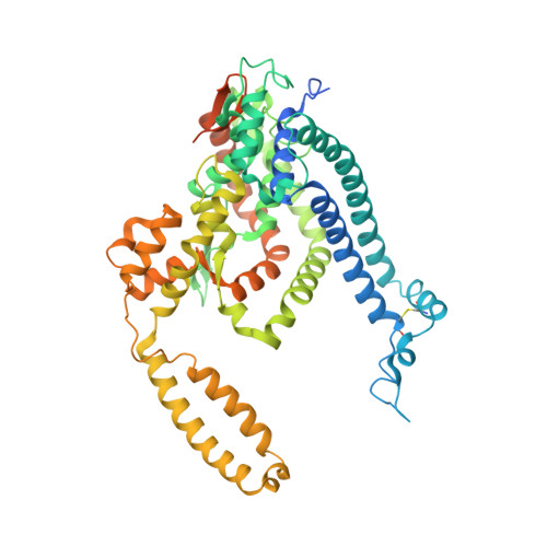
Structure, lipid scrambling activity and role in autophagosome formation of ATG9A.

Maeda, S.Yamamoto, H.Kinch, L.N.Garza, C.M.Takahashi, S.Otomo, C.Grishin, N.V.Forli, S.Mizushima, N.Otomo, T.

(2020) Nat Struct Mol Biol 27: 1194-1201

  • DOI: https://doi.org/10.1038/s41594-020-00520-2
  • Primary Citation Related Structures: 
    7JLO, 7JLP, 7JLQ

  • PubMed Abstract: 

    De novo formation of the double-membrane compartment autophagosome is seeded by small vesicles carrying membrane protein autophagy-related 9 (ATG9), the function of which remains unknown. Here we find that ATG9A scrambles phospholipids of membranes in vitro. Cryo-EM structures of human ATG9A reveal a trimer with a solvated central pore, which is connected laterally to the cytosol through the cavity within each protomer. Similarities to ABC exporters suggest that ATG9A could be a transporter that uses the central pore to function. Moreover, molecular dynamics simulation suggests that the central pore opens laterally to accommodate lipid headgroups, thereby enabling lipids to flip. Mutations in the pore reduce scrambling activity and yield markedly smaller autophagosomes, indicating that lipid scrambling by ATG9A is essential for membrane expansion. We propose ATG9A acts as a membrane-embedded funnel to facilitate lipid flipping and to redistribute lipids added to the outer leaflet of ATG9 vesicles, thereby enabling growth into autophagosomes.


  • Organizational Affiliation
    • Department of Integrative Structural and Computational Biology, The Scripps Research Institute, La Jolla, CA, USA.

Macromolecule Content 

  • Total Structure Weight: 200.39 kDa 
  • Atom Count: 11,700 
  • Modeled Residue Count: 1,422 
  • Deposited Residue Count: 1,734 
  • Unique protein chains: 1

Macromolecules

Find similar proteins by:|  3D Structure
Entity ID: 1
MoleculeChains  Sequence LengthOrganismDetailsImage
Autophagy-related protein 9A
A, B, C
578Homo sapiensMutation(s): 0 
Gene Names: ATG9AAPG9L1
Membrane Entity: Yes 
UniProt & NIH Common Fund Data Resources
Find proteins for Q7Z3C6 (Homo sapiens)
Explore Q7Z3C6 
Go to UniProtKB:  Q7Z3C6
PHAROS:  Q7Z3C6
GTEx:  ENSG00000198925 
Entity Groups
Sequence Clusters30% Identity50% Identity70% Identity90% Identity95% Identity100% Identity
UniProt GroupQ7Z3C6
Sequence Annotations
Expand
Reference Sequence

Experimental Data & Validation

Experimental Data

  • Method: ELECTRON MICROSCOPY
  • Resolution: 4.00 Å
  • Aggregation State: PARTICLE 
  • Reconstruction Method: SINGLE PARTICLE 
EM Software:
TaskSoftware PackageVersion
RECONSTRUCTIONRELION
MODEL REFINEMENTPHENIX

Structure Validation

View Full Validation Report


Entry History 

& Funding Information

Deposition Data


Funding OrganizationLocationGrant Number
National Institutes of Health/National Institute of General Medical Sciences (NIH/NIGMS)United StatesGM092740

Revision History  (Full details and data files)

  • Version 1.0: 2020-10-28
    Type: Initial release
  • Version 1.1: 2020-11-11
    Changes: Database references
  • Version 1.2: 2020-12-16
    Changes: Database references
  • Version 1.3: 2024-10-23
    Changes: Data collection, Database references, Refinement description, Structure summary