7JH0 | pdb_00007jh0

Crystallographic structure of glyceraldehyde-3-phosphate dehydrogenase from Schistosoma mansoni


Experimental Data Snapshot

  • Method: X-RAY DIFFRACTION
  • Resolution: 2.51 Å
  • R-Value Free: 
    0.273 (Depositor), 0.285 (DCC) 
  • R-Value Work: 
    0.213 (Depositor), 0.225 (DCC) 
  • R-Value Observed: 
    0.216 (Depositor) 

Starting Model: experimental
View more details

wwPDB Validation 3D Report Full Report

Validation slider image for 7JH0

This is version 1.3 of the entry. See complete history

Literature

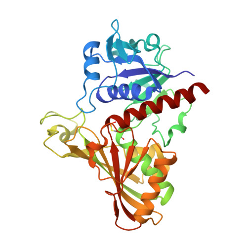
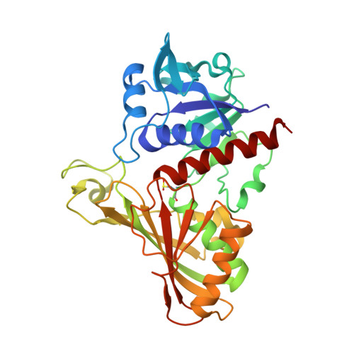
Structure determination and analyses of the GAPDH from the parasite Schistosoma mansoni, the first one from a platyhelminth.

Boreiko, S.Silva, M.Iulek, J.

(2021) Biochimie 184: 18-25

  • DOI: https://doi.org/10.1016/j.biochi.2021.01.014
  • Primary Citation Related Structures: 
    7JH0

  • PubMed Abstract: 

    The enzyme Glyceraldehyde-3-Phosphate Dehydrogenase from Schistosoma mansoni (SmGAPDH) is characterized as a therapeutical target for schistosomiasis. In this context, we report here the experimental structure, structural analyses and comparisons of SmGAPDH, the first one from a Platyhelminth. The enzyme was expressed, purified and assayed for crystallization, what allowed the obtainment of crystals of sufficient quality to collect X-ray diffraction data up to 2.51 Å resolution. SmGAPDH is the only GAPDH to present the sequence NNR (its residues 114-116) which leads to (especially R116) a hydrogen bond network that possibly reflects on the flexibility of residues to interact with the adenine part of NAD + , speculated to be important for differential drug design.


  • Organizational Affiliation
    • Department of Chemistry, State University of Ponta Grossa, Ponta Grossa - PR, 84030-900, Brazil.

Macromolecule Content 

  • Total Structure Weight: 146.73 kDa 
  • Atom Count: 10,188 
  • Modeled Residue Count: 1,352 
  • Deposited Residue Count: 1,352 
  • Unique protein chains: 2

Macromolecules

Find similar proteins by:|  3D Structure
Entity ID: 1
MoleculeChains  Sequence LengthOrganismDetailsImage
Glyceraldehyde-3-phosphate dehydrogenase
A, B, C
338Schistosoma mansoniMutation(s): 0 
EC: 1.2.1.12
UniProt
Find proteins for P20287 (Schistosoma mansoni)
Explore P20287 
Go to UniProtKB:  P20287
Entity Groups
Sequence Clusters30% Identity50% Identity70% Identity90% Identity95% Identity100% Identity
UniProt GroupP20287
Sequence Annotations
Expand
Reference Sequence
Find similar proteins by:|  3D Structure
Entity ID: 2
MoleculeChains  Sequence LengthOrganismDetailsImage
Glyceraldehyde-3-phosphate dehydrogenase338Schistosoma mansoniMutation(s): 0 
EC: 1.2.1.12
UniProt
Find proteins for P20287 (Schistosoma mansoni)
Explore P20287 
Go to UniProtKB:  P20287
Entity Groups
Sequence Clusters30% Identity50% Identity70% Identity90% Identity95% Identity100% Identity
UniProt GroupP20287
Sequence Annotations
Expand
Reference Sequence

Small Molecules

Ligands 4 Unique
IDChains Name / Formula / InChI Key2D Diagram3D Interactions
PGE

Query on PGE


Download:Ideal Coordinates CCD File
F [auth A],
I [auth B]
TRIETHYLENE GLYCOL
C6 H14 O4
ZIBGPFATKBEMQZ-UHFFFAOYSA-N
PEG

Query on PEG


Download:Ideal Coordinates CCD File
H [auth B],
K [auth D]
DI(HYDROXYETHYL)ETHER
C4 H10 O3
MTHSVFCYNBDYFN-UHFFFAOYSA-N
GOL

Query on GOL


Download:Ideal Coordinates CCD File
E [auth A],
G [auth B]
GLYCEROL
C3 H8 O3
PEDCQBHIVMGVHV-UHFFFAOYSA-N
EDO

Query on EDO


Download:Ideal Coordinates CCD File
J [auth C]1,2-ETHANEDIOL
C2 H6 O2
LYCAIKOWRPUZTN-UHFFFAOYSA-N
Modified Residues  2 Unique
IDChains TypeFormula2D DiagramParent
CSP
Query on CSP
A, B, C
L-PEPTIDE LINKINGC3 H8 N O5 P SCYS
CSD
Query on CSD
D
L-PEPTIDE LINKINGC3 H7 N O4 SCYS

Experimental Data & Validation

Experimental Data

  • Method: X-RAY DIFFRACTION
  • Resolution: 2.51 Å
  • R-Value Free:  0.273 (Depositor), 0.285 (DCC) 
  • R-Value Work:  0.213 (Depositor), 0.225 (DCC) 
  • R-Value Observed: 0.216 (Depositor) 
Space Group: P 61
Unit Cell:
Length ( Å )Angle ( ˚ )
a = 80.123α = 90
b = 80.123β = 90
c = 417.015γ = 120
Software Package:
Software NamePurpose
PHENIXrefinement
XSCALEdata scaling
XDSdata processing
PHENIXmodel building
PHASERphasing
XDSdata reduction

Structure Validation

View Full Validation Report


Entry History 

Deposition Data

Revision History  (Full details and data files)

  • Version 1.0: 2021-02-17
    Type: Initial release
  • Version 1.1: 2021-02-24
    Changes: Database references
  • Version 1.2: 2023-10-18
    Changes: Data collection, Database references, Refinement description
  • Version 1.3: 2024-11-20
    Changes: Structure summary