7H5C | pdb_00007h5c

Crystal structure of endothiapepsin PN_cryo3 in complex with AC40075 at 100 K


Experimental Data Snapshot

  • Method: X-RAY DIFFRACTION
  • Resolution: 1.91 Å
  • R-Value Free: 
    0.199 (Depositor), 0.195 (DCC) 
  • R-Value Work: 
    0.153 (Depositor), 0.153 (DCC) 
  • R-Value Observed: 
    0.156 (Depositor) 

Starting Model: experimental
View more details

wwPDB Validation 3D Report Full Report

Validation slider image for 7H5C

Ligand Structure Quality Assessment 


This is version 1.2 of the entry. See complete history

Literature

Cryo2RT: a high-throughput method for room-temperature macromolecular crystallography from cryo-cooled crystals.

Huang, C.Y.Aumonier, S.Olieric, V.Wang, M.

(2024) Acta Crystallogr D Struct Biol 80: 620-628

  • DOI: https://doi.org/10.1107/S2059798324006697
  • Primary Citation Related Structures: 
    7H56, 7H57, 7H58, 7H59, 7H5A, 7H5B, 7H5C, 7H5D, 7H5E, 7H5F, 7H5G, 7H5H, 7H5I, 7H5J, 7H5K, 7H5L, 7H5M, 7H5N, 7H5O, 7H5P, 7H5Q, 7H5R, 7H5S, 7H5T, 7H5U, 7H5V, 7H5W, 7H5X, 7H5Y, 7H5Z, 9FX4, 9FX5, 9FX6, 9FX7

  • PubMed Abstract: 

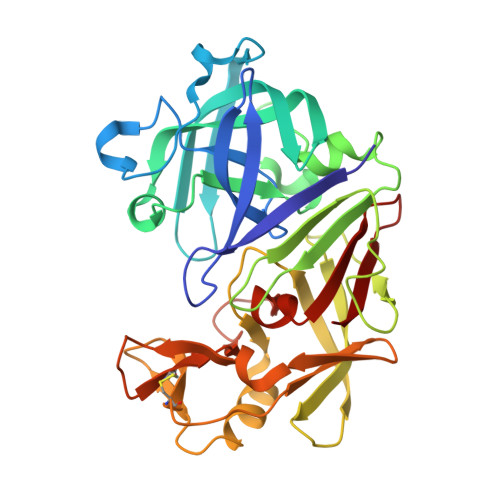
    Advances in structural biology have relied heavily on synchrotron cryo-crystallography and cryogenic electron microscopy to elucidate biological processes and for drug discovery. However, disparities between cryogenic and room-temperature (RT) crystal structures pose challenges. Here, Cryo2RT, a high-throughput RT data-collection method from frozen crystals that leverages the cryo-crystallography workflow, is introduced. Tested on endothiapepsin crystals with four soaked fragments, thaumatin and SARS-CoV-2 3CL pro , Cryo2RT reveals unique ligand-binding poses, offers a comparable throughput to cryo-crystallography and eases the exploration of structural dynamics at various temperatures.


  • Organizational Affiliation
    • Swiss Light Source, Center for Photon Science, Paul Scherrer Institute, Forschungsstrasse 111, 5232 Villigen PSI, Switzerland.

Macromolecule Content 

  • Total Structure Weight: 38.22 kDa 
  • Atom Count: 2,934 
  • Modeled Residue Count: 330 
  • Deposited Residue Count: 330 
  • Unique protein chains: 1

Macromolecules

Find similar proteins by:|  3D Structure
Entity ID: 1
MoleculeChains  Sequence LengthOrganismDetailsImage
Endothiapepsin330Cryphonectria parasiticaMutation(s): 0 
EC: 3.4.23.22
UniProt
Find proteins for P11838 (Cryphonectria parasitica)
Explore P11838 
Go to UniProtKB:  P11838
Entity Groups
Sequence Clusters30% Identity50% Identity70% Identity90% Identity95% Identity100% Identity
UniProt GroupP11838
Sequence Annotations
Expand
Reference Sequence

Small Molecules

Ligands 3 Unique
IDChains Name / Formula / InChI Key2D Diagram3D Interactions
EMR
(Subject of Investigation/LOI)

Query on EMR


Download:Ideal Coordinates CCD File
G [auth A]N-AMINOETHYLMORPHOLINE
C6 H14 N2 O
RWIVICVCHVMHMU-UHFFFAOYSA-N
DMS

Query on DMS


Download:Ideal Coordinates CCD File
AA [auth A]
AB [auth A]
BA [auth A]
BB [auth A]
C [auth A]
AA [auth A],
AB [auth A],
BA [auth A],
BB [auth A],
C [auth A],
CA [auth A],
CB [auth A],
D [auth A],
DA [auth A],
DB [auth A],
E [auth A],
EA [auth A],
EB [auth A],
F [auth A],
FA [auth A],
GA [auth A],
H [auth A],
HA [auth A],
I [auth A],
IA [auth A],
J [auth A],
JA [auth A],
K [auth A],
KA [auth A],
L [auth A],
LA [auth A],
M [auth A],
MA [auth A],
N [auth A],
NA [auth A],
O [auth A],
OA [auth A],
P [auth A],
PA [auth A],
Q [auth A],
QA [auth A],
R [auth A],
RA [auth A],
S [auth A],
SA [auth A],
T [auth A],
TA [auth A],
U [auth A],
UA [auth A],
V [auth A],
VA [auth A],
W [auth A],
WA [auth A],
X [auth A],
XA [auth A],
Y [auth A],
YA [auth A],
Z [auth A],
ZA [auth A]
DIMETHYL SULFOXIDE
C2 H6 O S
IAZDPXIOMUYVGZ-UHFFFAOYSA-N
ACT

Query on ACT


Download:Ideal Coordinates CCD File
B [auth A]ACETATE ION
C2 H3 O2
QTBSBXVTEAMEQO-UHFFFAOYSA-M

Experimental Data & Validation

Experimental Data

  • Method: X-RAY DIFFRACTION
  • Resolution: 1.91 Å
  • R-Value Free:  0.199 (Depositor), 0.195 (DCC) 
  • R-Value Work:  0.153 (Depositor), 0.153 (DCC) 
  • R-Value Observed: 0.156 (Depositor) 
Space Group: P 1 21 1
Unit Cell:
Length ( Å )Angle ( ˚ )
a = 43.464α = 90
b = 71.936β = 107.36
c = 50.85γ = 90
Software Package:
Software NamePurpose
BUSTERrefinement
XSCALEdata scaling
XDSdata reduction
PHASERphasing
PDB_EXTRACTdata extraction

Structure Validation

View Full Validation Report


Ligand Structure Quality Assessment 


Entry History 

& Funding Information

Deposition Data


Funding OrganizationLocationGrant Number
Swiss National Science FoundationSwitzerland182369
H2020 Marie Curie Actions of the European CommissionEuropean Union884104

Revision History  (Full details and data files)

  • Version 1.0: 2024-08-07
    Type: Initial release
  • Version 1.1: 2024-08-21
    Changes: Database references
  • Version 1.2: 2024-11-13
    Changes: Structure summary