7FSB | pdb_00007fsb

Structure of liver pyruvate kinase in complex with allosteric modulator 41


Experimental Data Snapshot

  • Method: X-RAY DIFFRACTION
  • Resolution: 2.50 Å
  • R-Value Free: 
    0.292 (Depositor), 0.275 (DCC) 
  • R-Value Work: 
    0.267 (Depositor), 0.258 (DCC) 
  • R-Value Observed: 
    0.268 (Depositor) 

Starting Model: experimental
View more details

wwPDB Validation 3D Report Full Report

Validation slider image for 7FSB

Ligand Structure Quality Assessment 


This is version 1.1 of the entry. See complete history

Literature

Tuning liver pyruvate kinase activity up or down with a new class of allosteric modulators.

Nain-Perez, A.Nilsson, O.Lulla, A.Haversen, L.Brear, P.Liljenberg, S.Hyvonen, M.Boren, J.Grotli, M.

(2023) Eur J Med Chem 250: 115177-115177

  • DOI: https://doi.org/10.1016/j.ejmech.2023.115177
  • Primary Citation Related Structures: 
    7FRV, 7FRW, 7FRX, 7FRY, 7FRZ, 7FS0, 7FS1, 7FS2, 7FS3, 7FS4, 7FS5, 7FS6, 7FS7, 7FS8, 7FS9, 7FSA, 7FSB, 7FSC, 7FSD

  • PubMed Abstract: 

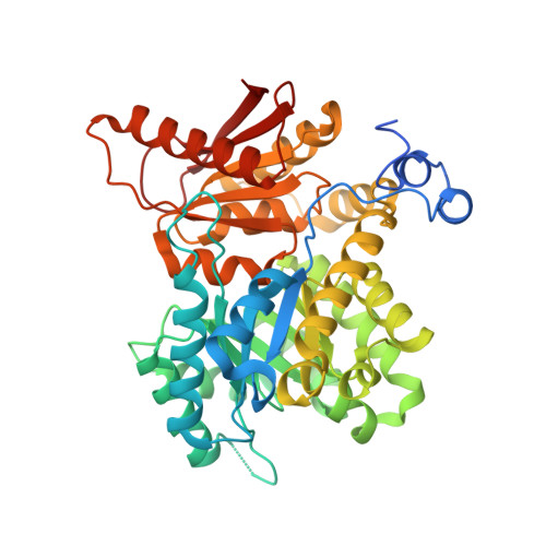
    The liver isoform of pyruvate kinase (PKL) has gained interest due to its potential capacity to regulate fatty acid synthesis involved in the progression of non-alcoholic fatty liver disease (NAFLD). Here we describe a novel series of PKL modulators that can either activate or inhibit the enzyme allosterically, from a cryptic site at the interface of two protomers in the tetrameric enzyme. Starting from urolithin D, we designed and synthesised 42 new compounds. The effect of these compounds on PKL enzymatic activity was assessed after incubation with cell lysates obtained from a liver cell line. Pronounced activation of PKL activity, up to 3.8-fold, was observed for several compounds at 10 μM, while other compounds were prominent PKL inhibitors reducing its activity to 81% at best. A structure-activity relationship identified linear-shaped sulfone-sulfonamides as activators and non-linear compounds as inhibitors. Crystal structures revealed the conformations of these modulators, which were used as a reference for designing new modulators.


  • Organizational Affiliation
    • Department of Chemistry and Molecular Biology, University of Gothenburg, SE-412 96, Gothenburg, Sweden.

Macromolecule Content 

  • Total Structure Weight: 391.82 kDa 
  • Atom Count: 26,850 
  • Modeled Residue Count: 3,405 
  • Deposited Residue Count: 3,576 
  • Unique protein chains: 1

Macromolecules

Find similar proteins by:|  3D Structure
Entity ID: 1
MoleculeChains  Sequence LengthOrganismDetailsImage
Pyruvate kinase PKLR
A, B, C, D, E
A, B, C, D, E, F, G, H
447Homo sapiensMutation(s): 0 
Gene Names: PKLRPK1PKL
EC: 2.7.1.40
UniProt & NIH Common Fund Data Resources
Find proteins for P30613 (Homo sapiens)
Explore P30613 
Go to UniProtKB:  P30613
PHAROS:  P30613
GTEx:  ENSG00000143627 
Entity Groups
Sequence Clusters30% Identity50% Identity70% Identity90% Identity95% Identity100% Identity
UniProt GroupP30613
Sequence Annotations
Expand
Reference Sequence

Small Molecules

Ligands 5 Unique
IDChains Name / Formula / InChI Key2D Diagram3D Interactions
OE0
(Subject of Investigation/LOI)

Query on OE0


Download:Ideal Coordinates CCD File
IA [auth F],
M [auth A],
NA [auth G],
R [auth B]
N-({4-[(3,4-dihydroxyphenyl)methyl]phenyl}methyl)-3,4-dihydroxybenzene-1-sulfonamide
C20 H19 N O6 S
WNFFMCJBFDREPV-UHFFFAOYSA-N
FBP

Query on FBP


Download:Ideal Coordinates CCD File
AA [auth E]
EA [auth F]
I [auth A]
JA [auth G]
N [auth B]
AA [auth E],
EA [auth F],
I [auth A],
JA [auth G],
N [auth B],
OA [auth H],
S [auth C],
W [auth D]
1,6-di-O-phosphono-beta-D-fructofuranose
C6 H14 O12 P2
RNBGYGVWRKECFJ-ARQDHWQXSA-N
OXL

Query on OXL


Download:Ideal Coordinates CCD File
BA [auth E]
FA [auth F]
J [auth A]
KA [auth G]
O [auth B]
BA [auth E],
FA [auth F],
J [auth A],
KA [auth G],
O [auth B],
PA [auth H],
T [auth C],
X [auth D]
OXALATE ION
C2 O4
MUBZPKHOEPUJKR-UHFFFAOYSA-L
K

Query on K


Download:Ideal Coordinates CCD File
DA [auth E]
HA [auth F]
L [auth A]
MA [auth G]
Q [auth B]
DA [auth E],
HA [auth F],
L [auth A],
MA [auth G],
Q [auth B],
RA [auth H],
V [auth C],
Z [auth D]
POTASSIUM ION
K
NPYPAHLBTDXSSS-UHFFFAOYSA-N
MG

Query on MG


Download:Ideal Coordinates CCD File
CA [auth E]
GA [auth F]
K [auth A]
LA [auth G]
P [auth B]
CA [auth E],
GA [auth F],
K [auth A],
LA [auth G],
P [auth B],
QA [auth H],
U [auth C],
Y [auth D]
MAGNESIUM ION
Mg
JLVVSXFLKOJNIY-UHFFFAOYSA-N

Experimental Data & Validation

Experimental Data

  • Method: X-RAY DIFFRACTION
  • Resolution: 2.50 Å
  • R-Value Free:  0.292 (Depositor), 0.275 (DCC) 
  • R-Value Work:  0.267 (Depositor), 0.258 (DCC) 
  • R-Value Observed: 0.268 (Depositor) 
Space Group: C 1 2 1
Unit Cell:
Length ( Å )Angle ( ˚ )
a = 208.049α = 90
b = 113.231β = 91.84
c = 188.473γ = 90
Software Package:
Software NamePurpose
XDSdata reduction
PHASERphasing
BUSTERrefinement
PDB_EXTRACTdata extraction
STARANISOdata scaling

Structure Validation

View Full Validation Report


Ligand Structure Quality Assessment 


Entry History 

& Funding Information

Deposition Data


Funding OrganizationLocationGrant Number
Not fundedUnited Kingdom--

Revision History  (Full details and data files)

  • Version 1.0: 2023-04-12
    Type: Initial release
  • Version 1.1: 2024-05-22
    Changes: Data collection