


Crystal Structure of a human Legumain complex
Bartels, B., Kuhn, B., Benz, J., Rudolph, M.G.To be published.
Experimental Data Snapshot
Starting Model: other
View more details
Entity ID: 1 | |||||
|---|---|---|---|---|---|
| Molecule | Chains | Sequence Length | Organism | Details | Image |
| Legumain | A, B, C [auth D] | 444 | Homo sapiens | Mutation(s): 0 Gene Names: LGMN, PRSC1 EC: 3.4.22.34 | 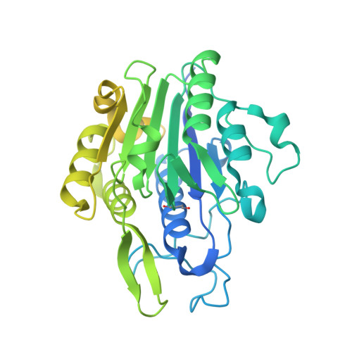 |
UniProt & NIH Common Fund Data Resources | |||||
PHAROS: Q99538 GTEx: ENSG00000100600 | |||||
Entity Groups | |||||
| Sequence Clusters | 30% Identity50% Identity70% Identity90% Identity95% Identity100% Identity | ||||
| UniProt Group | Q99538 | ||||
Glycosylation | |||||
| Glycosylation Sites: 1 | Go to GlyGen: Q99538-1 | ||||
Sequence AnnotationsExpand | |||||
Reference Sequence | |||||
| Ligands 3 Unique | |||||
|---|---|---|---|---|---|
| ID | Chains | Name / Formula / InChI Key | 2D Diagram | 3D Interactions | |
| WR9 (Subject of Investigation/LOI) Download:Ideal Coordinates CCD File | D [auth A], G [auth B], J [auth D] | N-[(3S)-5-amino-5-oxopent-1-en-3-yl]-1-{1-[4-(cyclopropylmethoxy)phenyl]cyclopropane-1-carbonyl}-L-prolinamide C24 H31 N3 O4 SOCJRKDNYUYZLL-QUCCMNQESA-N |  | ||
| NAG Download:Ideal Coordinates CCD File | F [auth A], H [auth B] | 2-acetamido-2-deoxy-beta-D-glucopyranose C8 H15 N O6 OVRNDRQMDRJTHS-FMDGEEDCSA-N |  | ||
| EDO Download:Ideal Coordinates CCD File | E [auth A], I [auth B] | 1,2-ETHANEDIOL C2 H6 O2 LYCAIKOWRPUZTN-UHFFFAOYSA-N |  | ||
| Modified Residues 1 Unique | |||||
|---|---|---|---|---|---|
| ID | Chains | Type | Formula | 2D Diagram | Parent |
| SNN Query on SNN | A, B, C [auth D] | L-PEPTIDE LINKING | C4 H6 N2 O2 |  | ASN |
| Length ( Å ) | Angle ( ˚ ) |
|---|---|
| a = 117.798 | α = 90 |
| b = 117.798 | β = 90 |
| c = 102.812 | γ = 120 |
| Software Name | Purpose |
|---|---|
| XSCALE | data scaling |
| REFMAC | refinement |
| PDB_EXTRACT | data extraction |
| XDS | data reduction |
| XSCALE | data scaling |
| PHASER | phasing |
| Funding Organization | Location | Grant Number |
|---|---|---|
| F. Hoffmann-La Roche | Switzerland | -- |


