7FET | pdb_00007fet

SARS-CoV-2 B.1.1.7 Spike Glycoprotein trimer


Experimental Data Snapshot

  • Method: ELECTRON MICROSCOPY
  • Resolution: 3.70 Å
  • Aggregation State: PARTICLE 
  • Reconstruction Method: SINGLE PARTICLE 

wwPDB Validation 3D Report Full Report

Validation slider image for 7FET

This is version 1.1 of the entry. See complete history

Literature

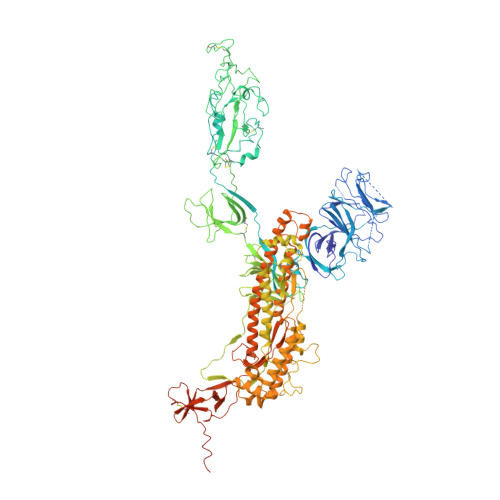
Structure-based evidence for the enhanced transmissibility of the dominant SARS-CoV-2 B.1.1.7 variant (Alpha).

Xia, S.Wen, Z.Wang, L.Lan, Q.Jiao, F.Tai, L.Wang, Q.Sun, F.Jiang, S.Lu, L.Zhu, Y.

(2021) Cell Discov 7: 109-109

  • DOI: https://doi.org/10.1038/s41421-021-00349-z
  • Primary Citation Related Structures: 
    7FEM, 7FET

  • Organizational Affiliation
    • Key Laboratory of Medical Molecular Virology (MOE/NHC/CAMS), School of Basic Medical Sciences and Biosafety Level 3 Laboratory, Shanghai Institute of Infectious Disease and Biosecurity, Fudan University, Shanghai, China.

Macromolecule Content 

  • Total Structure Weight: 404.53 kDa 
  • Atom Count: 24,023 
  • Modeled Residue Count: 3,003 
  • Deposited Residue Count: 3,573 
  • Unique protein chains: 1

Macromolecules

Find similar proteins by:|  3D Structure
Entity ID: 1
MoleculeChains  Sequence LengthOrganismDetailsImage
Spike glycoprotein
A, B, C
1,191Severe acute respiratory syndrome coronavirus 2Mutation(s): 12 
Gene Names: S2
UniProt
Find proteins for P0DTC2 (Severe acute respiratory syndrome coronavirus 2)
Explore P0DTC2 
Go to UniProtKB:  P0DTC2
Entity Groups
Sequence Clusters30% Identity50% Identity70% Identity90% Identity95% Identity100% Identity
UniProt GroupP0DTC2
Glycosylation
Glycosylation Sites: 10Go to GlyGen: P0DTC2-1
Sequence Annotations
Expand
Reference Sequence

Oligosaccharides

Help  
Entity ID: 2
MoleculeChains Length2D Diagram GlycosylationD Interactions
2-acetamido-2-deoxy-beta-D-glucopyranose-(1-4)-2-acetamido-2-deoxy-beta-D-glucopyranose
D [auth E],
E [auth F],
F [auth H],
G [auth I],
H [auth P],
D [auth E],
E [auth F],
F [auth H],
G [auth I],
H [auth P],
I [auth Q],
J [auth S],
K [auth U],
L [auth V]
2N-Glycosylation
Glycosylation Resources
GlyTouCan: G42666HT
GlyCosmos: G42666HT
GlyGen: G42666HT

Small Molecules

Ligands 1 Unique
IDChains Name / Formula / InChI Key2D Diagram3D Interactions
NAG

Query on NAG


Download:Ideal Coordinates CCD File
AA [auth B]
BA [auth B]
CA [auth B]
DA [auth C]
EA [auth C]
AA [auth B],
BA [auth B],
CA [auth B],
DA [auth C],
EA [auth C],
FA [auth C],
GA [auth C],
HA [auth C],
IA [auth C],
M [auth A],
N [auth A],
O [auth A],
P [auth A],
Q [auth A],
R [auth A],
S [auth A],
T [auth A],
U [auth B],
V [auth B],
W [auth B],
X [auth B],
Y [auth B],
Z [auth B]
2-acetamido-2-deoxy-beta-D-glucopyranose
C8 H15 N O6
OVRNDRQMDRJTHS-FMDGEEDCSA-N

Experimental Data & Validation

Experimental Data

  • Method: ELECTRON MICROSCOPY
  • Resolution: 3.70 Å
  • Aggregation State: PARTICLE 
  • Reconstruction Method: SINGLE PARTICLE 

Structure Validation

View Full Validation Report


Entry History 

& Funding Information

Deposition Data


Funding OrganizationLocationGrant Number
Not funded--

Revision History  (Full details and data files)

  • Version 1.0: 2021-12-15
    Type: Initial release
  • Version 1.1: 2024-10-23
    Changes: Data collection, Structure summary