A cryptic-site ligand stabilizes a non-canonical interface and blocks membrane insertion of the chloride intracellular channel CLIC1.
Kuila, S., Ghoshal, A., Sahoo, S., Aathi, M., Khan, M.A., Panchariya, L., Sonkar, K.S., Khan, W.A., Pandiramesh, J.R., Chandele, A., Arulandu, A.(2026) J Biological Chem 302: 113113-113113
- PubMed: 42103216 Search on PubMedSearch on PubMed Central
- DOI: https://doi.org/10.1016/j.jbc.2026.113113
- Primary Citation Related Structures:
24UG, 7FBQ - PubMed Abstract:
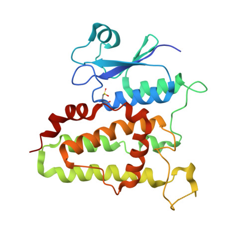
Human chloride intracellular channel 1 (HsCLIC1) is a dimorphic protein that exists in both soluble enzymatic and membrane-integrated ion channel forms. It is overexpressed in many cancers, yet developing specific inhibitors has been challenging, leaving CLIC proteins largely untargeted. Here, we identified a non-canonical ligand binding site on the soluble enzymatic form of HsCLIC1 and demonstrate that it is druggable with the small molecule inhibitor NSC602247. This cryptic site was discovered using multi-solvent molecular dynamics (MD) simulations followed by virtual screening and molecular docking. Using surface plasmon resonance and microscale thermophoresis, we show that NSC602247 binds to HsCLIC1 with micromolar affinity (∼6 μM) and inhibits the proliferation of HT29 colorectal cancer cells. To understand the mechanism of inhibition, we determined the crystal structure of the HsCLIC1-NSC602247 complex. Mechanistically, NSC602247 binding induces oligomerization of soluble HsCLIC1 and reduces its membrane expression, as revealed using flow cytometry. Our data clearly indicate that NSC602247 inhibits HT29 growth by sequestering the protein in a soluble state, thereby blocking its membrane translocation. These findings validate a novel cryptic site on CLIC1 and present a new mechanistic paradigm for inhibiting its pathological function by preventing membrane insertion, offering a promising strategy for targeted therapeutic development.
- Structural Biology Group, International Centre for Genetic Engineering and Biotechnology, New Delhi, India.
Organizational Affiliation: