7FB7

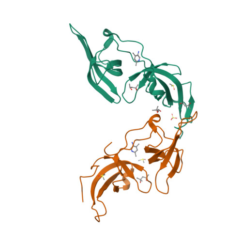
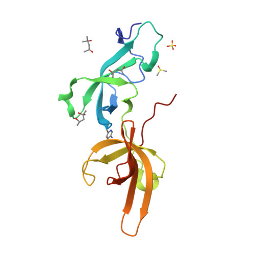
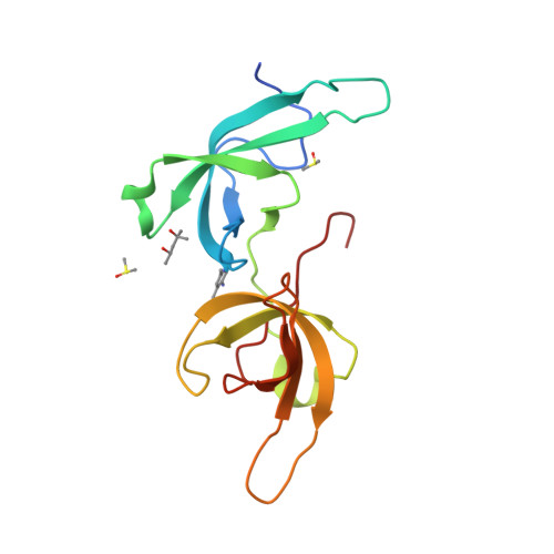
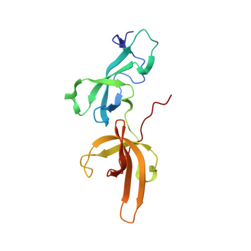
Crystal structure of human UHRF1 TTD in complex with 5-amino-2,4-dimethylpyridine


Experimental Data Snapshot

  • Method: X-RAY DIFFRACTION
  • Resolution: 1.45 Å
  • R-Value Free: 0.173 
  • R-Value Work: 0.158 
  • R-Value Observed: 0.158 

Starting Model: experimental
View more details

wwPDB Validation   3D Report Full Report


Ligand Structure Quality Assessment 


This is version 1.1 of the entry. See complete history


Literature

Structure-based screening combined with computational and biochemical analyses identified the inhibitor targeting the binding of DNA Ligase 1 to UHRF1.

Kori, S.Shibahashi, Y.Ekimoto, T.Nishiyama, A.Yoshimi, S.Yamaguchi, K.Nagatoishi, S.Ohta, M.Tsumoto, K.Nakanishi, M.Defossez, P.A.Ikeguchi, M.Arita, K.

(2021) Bioorg Med Chem 52: 116500-116500

  • DOI: https://doi.org/10.1016/j.bmc.2021.116500
  • Primary Citation of Related Structures:  
    7FB7

  • PubMed Abstract: 

    The accumulation of epigenetic alterations is one of the major causes of tumorigenesis. Aberrant DNA methylation patterns cause genome instability and silencing of tumor suppressor genes in various types of tumors. Therefore, drugs that target DNA methylation-regulating factors have great potential for cancer therapy. Ubiquitin-like containing PHD and RING finger domain 1 (UHRF1) is an essential factor for DNA methylation maintenance. UHRF1 is overexpressed in various cancer cells and down-regulation of UHRF1 in these cells reactivates the expression of tumor suppressor genes, thus UHRF1 is a promising target for cancer therapy. We have previously shown that interaction between the tandem Tudor domain (TTD) of UHRF1 and DNA ligase 1 (LIG1) di/trimethylated on Lys126 plays a key role in the recruitment of UHRF1 to replication sites and replication-coupled DNA methylation maintenance. An arginine binding cavity (Arg-binding cavity) of the TTD is essential for LIG1 interaction, thus the development of inhibitors that target the Arg-binding cavity could potentially repress UHRF1 function in cancer cells. To develop such an inhibitor, we performed in silico screening using not only static but also dynamic metrics based on all-atom molecular dynamics simulations, resulting in efficient identification of 5-amino-2,4-dimethylpyridine (5A-DMP) as a novel TTD-binding compound. Crystal structure of the TTD in complex with 5A-DMP revealed that the compound stably bound to the Arg-binding cavity of the TTD. Furthermore, 5A-DMP inhibits the full-length UHRF1:LIG1 interaction in Xenopus egg extracts. Our study uncovers a UHRF1 inhibitor which can be the basis of future experiments for cancer therapy.


  • Organizational Affiliation

    Structural Biology Laboratory, Graduate School of Medical Life Science, Yokohama City University, 1-7-29, Suehiro-cho, Tsurumi-ku, Yokohama, Kanagawa 230-0045, Japan.


Macromolecules
Find similar proteins by:  (by identity cutoff)  |  3D Structure
Entity ID: 1
MoleculeChains Sequence LengthOrganismDetailsImage
E3 ubiquitin-protein ligase UHRF1
A, B
155Homo sapiensMutation(s): 0 
Gene Names: UHRF1ICBP90NP95RNF106
EC: 2.3.2.27
UniProt & NIH Common Fund Data Resources
Find proteins for Q96T88 (Homo sapiens)
Explore Q96T88 
Go to UniProtKB:  Q96T88
PHAROS:  Q96T88
GTEx:  ENSG00000276043 
Entity Groups  
Sequence Clusters30% Identity50% Identity70% Identity90% Identity95% Identity100% Identity
UniProt GroupQ96T88
Sequence Annotations
Expand
  • Reference Sequence
Small Molecules
Ligands 4 Unique
IDChains Name / Formula / InChI Key2D Diagram3D Interactions
8NF (Subject of Investigation/LOI)
Query on 8NF

Download Ideal Coordinates CCD File 
F [auth A],
J [auth B]
5-amino-2,4-dimethylpyridine
C7 H10 N2
SHSQTJRJQHPJRQ-UHFFFAOYSA-N
MPD
Query on MPD

Download Ideal Coordinates CCD File 
C [auth A],
D [auth A],
E [auth A],
I [auth B]
(4S)-2-METHYL-2,4-PENTANEDIOL
C6 H14 O2
SVTBMSDMJJWYQN-YFKPBYRVSA-N
SO4
Query on SO4

Download Ideal Coordinates CCD File 
H [auth A]SULFATE ION
O4 S
QAOWNCQODCNURD-UHFFFAOYSA-L
DMS
Query on DMS

Download Ideal Coordinates CCD File 
G [auth A],
K [auth B],
L [auth B]
DIMETHYL SULFOXIDE
C2 H6 O S
IAZDPXIOMUYVGZ-UHFFFAOYSA-N
Binding Affinity Annotations 
IDSourceBinding Affinity
8NF BindingDB:  7FB7 Kd: min: 1.93e+4, max: 1.83e+5 (nM) from 5 assay(s)
Experimental Data & Validation

Experimental Data

  • Method: X-RAY DIFFRACTION
  • Resolution: 1.45 Å
  • R-Value Free: 0.173 
  • R-Value Work: 0.158 
  • R-Value Observed: 0.158 
  • Space Group: H 3 2
Unit Cell:
Length ( Å )Angle ( ˚ )
a = 147.12α = 90
b = 147.12β = 90
c = 110.44γ = 120
Software Package:
Software NamePurpose
XDSdata scaling
PHENIXrefinement
Cootmodel building
Aimlessdata scaling
PHASERphasing

Structure Validation

View Full Validation Report


Ligand Structure Quality Assessment 


Entry History & Funding Information

Deposition Data


Funding OrganizationLocationGrant Number
Japan Society for the Promotion of Science (JSPS)JapanJP18H02392
Japan Society for the Promotion of Science (JSPS)JapanJP19H05294
Japan Society for the Promotion of Science (JSPS)JapanJP19H05741
Japan Society for the Promotion of Science (JSPS)JapanJP19J22030

Revision History  (Full details and data files)

  • Version 1.0: 2022-01-05
    Type: Initial release
  • Version 1.1: 2023-11-29
    Changes: Data collection, Refinement description