7FAB | pdb_00007fab

CRYSTAL STRUCTURE OF HUMAN IMMUNOGLOBULIN FRAGMENT FAB NEW REFINED AT 2.0 ANGSTROMS RESOLUTION


Experimental Data Snapshot

  • Method: X-RAY DIFFRACTION
  • Resolution: 2.00 Å
  • R-Value Work: 
    0.169 (Depositor) 
  • R-Value Observed: 
    0.169 (Depositor) 

wwPDB Validation 3D Report Full Report

Validation slider image for 7FAB

This is version 1.5 of the entry. See complete history

Literature

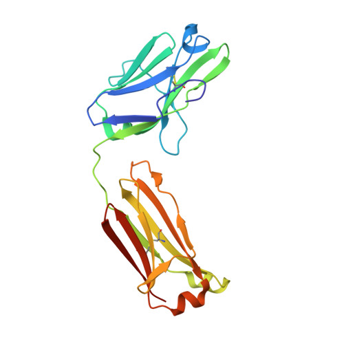
Crystal structure of human immunoglobulin fragment Fab New refined at 2.0 A resolution.

Saul, F.A.Poljak, R.J.

(1992) Proteins 14: 363-371

  • DOI: https://doi.org/10.1002/prot.340140305
  • Primary Citation Related Structures: 
    7FAB

  • PubMed Abstract: 

    The three-dimensional structure of the human immunoglobulin fragment Fab New (IgG1, lambda) has been refined to a crystallographic R-factor of 16.9% to 2 A resolution. Rms deviations of the final model from ideal geometry are 0.014 A for bond distances and 3.03 degrees for bond angles. Refinement was based on a new X-ray data set including 28,301 reflections with F > 2.5 sigma(F) from 6.0 to 2.0 A resolution. The starting model for the refinement procedure reported here is from the Brookhaven Protein Data Bank entry 3FAB (rev. 1981). Differences between the initial and final models include modified polypeptide-chain folding in the third complementarity-determining region (CDR3) and the third framework region (FR3) of VH and in some exposed loops of CL and CH1. Amino acid sequence changes were determined at a number of positions by inspection of difference electron density maps. The incorporation of amino acid sequence changes results in an improved VH framework model for the "humanization" of monoclonal antibodies.


  • Organizational Affiliation
    • Département d'Immunologie, Institut Pasteur, Paris, France.

Macromolecule Content 

  • Total Structure Weight: 45.06 kDa 
  • Atom Count: 3,313 
  • Modeled Residue Count: 413 
  • Deposited Residue Count: 425 
  • Unique protein chains: 2

Macromolecules

Find similar proteins by:|  3D Structure
Entity ID: 1
MoleculeChains  Sequence LengthOrganismDetailsImage
IGG1-LAMBDA NEW FAB (LIGHT CHAIN)A [auth L]208Homo sapiensMutation(s): 0 
UniProt & NIH Common Fund Data Resources
Find proteins for P0DOY2 (Homo sapiens)
Explore P0DOY2 
Go to UniProtKB:  P0DOY2
GTEx:  ENSG00000211677 
Entity Groups
Sequence Clusters30% Identity50% Identity70% Identity90% Identity95% Identity100% Identity
UniProt GroupP0DOY2
Sequence Annotations
Expand
Reference Sequence
Find similar proteins by:|  3D Structure
Entity ID: 2
MoleculeChains  Sequence LengthOrganismDetailsImage
IGG1-LAMBDA NEW FAB (HEAVY CHAIN)B [auth H]217Homo sapiensMutation(s): 0 
UniProt & NIH Common Fund Data Resources
Find proteins for P01825 (Homo sapiens)
Explore P01825 
Go to UniProtKB:  P01825
GTEx:  ENSG00000224373 
Entity Groups
Sequence Clusters30% Identity50% Identity70% Identity90% Identity95% Identity100% Identity
UniProt GroupP01825
Sequence Annotations
Expand
Reference Sequence

Experimental Data & Validation

Experimental Data

  • Method: X-RAY DIFFRACTION
  • Resolution: 2.00 Å
  • R-Value Work:  0.169 (Depositor) 
  • R-Value Observed: 0.169 (Depositor) 
Space Group: C 1 2 1
Unit Cell:
Length ( Å )Angle ( ˚ )
a = 111.43α = 90
b = 56.68β = 116.46
c = 90.3γ = 90
Software Package:
Software NamePurpose
X-PLORmodel building
X-PLORrefinement
X-PLORphasing

Structure Validation

View Full Validation Report


Entry History 

Deposition Data

Revision History  (Full details and data files)

  • Version 1.0: 1994-01-31
    Type: Initial release
  • Version 1.1: 2008-03-25
    Changes: Version format compliance
  • Version 1.2: 2011-07-13
    Changes: Version format compliance
  • Version 1.3: 2013-07-24
    Changes: Database references, Structure summary
  • Version 1.4: 2024-06-05
    Changes: Data collection, Database references, Other
  • Version 1.5: 2024-10-16
    Changes: Structure summary