7F86

Crystal structure of Phycoerythrin from Halomicronema Sp. R31DM


Experimental Data Snapshot

  • Method: X-RAY DIFFRACTION
  • Resolution: 2.21 Å
  • R-Value Free: 0.210 
  • R-Value Work: 0.163 
  • R-Value Observed: 0.165 

Starting Model: experimental
View more details

wwPDB Validation   3D Report Full Report


Ligand Structure Quality Assessment 


This is version 1.2 of the entry. See complete history


Literature

Crystal structure analysis of phycoerythrin from marine cyanobacterium Halomicronema .

Patel, S.N.Sonani, R.R.Gupta, G.D.Singh, N.K.Kumar, V.Madamwar, D.

(2023) J Biomol Struct Dyn 41: 3752-3761

  • DOI: https://doi.org/10.1080/07391102.2022.2055647
  • Primary Citation of Related Structures:  
    7F86

  • PubMed Abstract: 

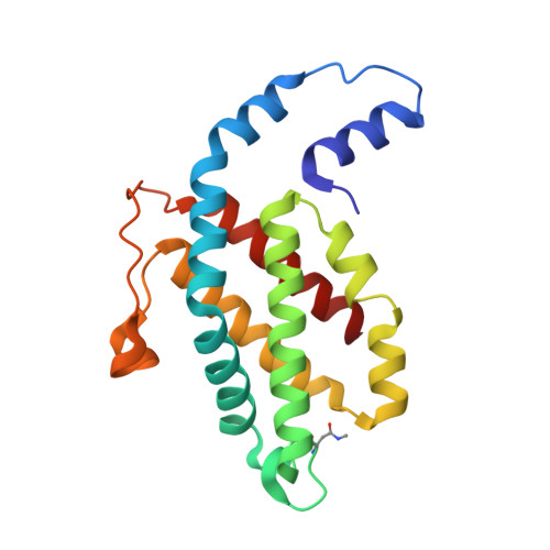
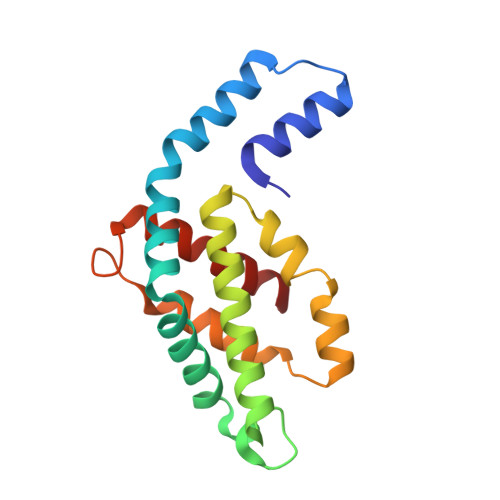
    Phycoerythrin (PE) is green light-absorbing pigment-protein that assists in efficient light harvesting in cyanobacteria and red-algae. PE in cyanobacteria stays less studied so far as compared to that in red algae. In this study, PE from marine cyanobacteria Halomicronema sp. R31DM is purified and subjected for its structural characterisation by X-ray crystallography in order to understand its light-harvesting characteristics. The crystal structure is solved to a resolution-limit of 2.21 Å with reasonable R-factors values, 0.16/0.21 (R work / R free ). PE forms hexamer of hetero-dimers made up of two peptide chains, α- and β-subunits containing 2 and 3 phycoerythrobilin (PEB) chromophores covalently attached to them, respectively. Geometry of five chromophores is analysed along with their relative position within the PE hexamer. Also, their interactions with the surrounding microenvironment are analysed. The plausible energy transfer pathways in hexamer structure have been predicted based on relative position and geometry of chromophores. This structure enriches the structural information of cyanobacterial PE in order to understand its light-harvesting capacity.Communicated by Ramaswamy H. Sarma.


  • Organizational Affiliation

    P. D. Patel Institute of Applied Sciences, Charotar University of Science and Technology, Anand, India.


Macromolecules
Find similar proteins by:  (by identity cutoff)  |  3D Structure
Entity ID: 1
MoleculeChains Sequence LengthOrganismDetailsImage
Phycoerythrin beta subunit184Halomicronema sp. R31DMMutation(s): 0 
UniProt
Find proteins for A0A1W1IW87 (Halomicronema sp. R31DM)
Explore A0A1W1IW87 
Go to UniProtKB:  A0A1W1IW87
Entity Groups  
Sequence Clusters30% Identity50% Identity70% Identity90% Identity95% Identity100% Identity
UniProt GroupA0A1W1IW87
Sequence Annotations
Expand
  • Reference Sequence
Find similar proteins by:  (by identity cutoff)  |  3D Structure
Entity ID: 2
MoleculeChains Sequence LengthOrganismDetailsImage
Phycoerythrin alpha subunit164Halomicronema sp. R31DMMutation(s): 0 
UniProt
Find proteins for A0A1W1IVY1 (Halomicronema sp. R31DM)
Explore A0A1W1IVY1 
Go to UniProtKB:  A0A1W1IVY1
Entity Groups  
Sequence Clusters30% Identity50% Identity70% Identity90% Identity95% Identity100% Identity
UniProt GroupA0A1W1IVY1
Sequence Annotations
Expand
  • Reference Sequence
Small Molecules
Ligands 1 Unique
IDChains Name / Formula / InChI Key2D Diagram3D Interactions
PEB (Subject of Investigation/LOI)
Query on PEB

Download Ideal Coordinates CCD File 
AA [auth M]
AB [auth E]
AC [auth V]
BA [auth A]
BB [auth E]
AA [auth M],
AB [auth E],
AC [auth V],
BA [auth A],
BB [auth E],
BC [auth X],
CA [auth A],
CB [auth E],
CC [auth X],
DA [auth D],
DB [auth G],
DC [auth X],
EA [auth D],
EB [auth G],
EC [auth Y],
FA [auth B],
FB [auth G],
FC [auth Y],
GA [auth B],
GB [auth I],
HA [auth F],
HB [auth I],
IA [auth F],
IB [auth I],
JA [auth H],
JB [auth K],
KA [auth H],
KB [auth K],
LA [auth J],
LB [auth K],
MA [auth J],
MB [auth N],
NA [auth L],
NB [auth N],
OA [auth L],
OB [auth N],
PA [auth O],
PB [auth P],
QA [auth O],
QB [auth P],
RA [auth Q],
RB [auth P],
SA [auth Q],
SB [auth R],
TA [auth U],
TB [auth R],
UA [auth U],
UB [auth R],
VA [auth W],
VB [auth T],
WA [auth W],
WB [auth T],
XA [auth C],
XB [auth T],
Y [auth M],
YA [auth C],
YB [auth V],
Z [auth M],
ZA [auth C],
ZB [auth V]
PHYCOERYTHROBILIN
C33 H40 N4 O6
NKCBCVIFPXGHAV-WAVSMFBNSA-N
Modified Residues  1 Unique
IDChains TypeFormula2D DiagramParent
MEN
Query on MEN
A [auth M]
M [auth C]
N [auth E]
O [auth G]
P [auth I]
A [auth M],
M [auth C],
N [auth E],
O [auth G],
P [auth I],
Q [auth K],
R [auth N],
S [auth P],
T [auth R],
U [auth T],
V,
W [auth X]
L-PEPTIDE LINKINGC5 H10 N2 O3ASN
Experimental Data & Validation

Experimental Data

  • Method: X-RAY DIFFRACTION
  • Resolution: 2.21 Å
  • R-Value Free: 0.210 
  • R-Value Work: 0.163 
  • R-Value Observed: 0.165 
  • Space Group: P 1
Unit Cell:
Length ( Å )Angle ( ˚ )
a = 108.59α = 78.37
b = 108.63β = 82.53
c = 116.93γ = 62.11
Software Package:
Software NamePurpose
REFMACrefinement
PDB_EXTRACTdata extraction
iMOSFLMdata reduction
Aimlessdata scaling
PHASERphasing

Structure Validation

View Full Validation Report


Ligand Structure Quality Assessment 


Entry History & Funding Information

Deposition Data


Funding OrganizationLocationGrant Number
Department of Biotechnology (DBT, India)IndiaBT/PR15686/AAQ/3/811/2016

Revision History  (Full details and data files)

  • Version 1.0: 2022-04-13
    Type: Initial release
  • Version 1.1: 2023-05-24
    Changes: Database references
  • Version 1.2: 2023-11-29
    Changes: Data collection, Refinement description