7F7E | pdb_00007f7e

SARS-CoV-2 S protein RBD in complex with A5-10 Fab


Experimental Data Snapshot

  • Method: X-RAY DIFFRACTION
  • Resolution: 2.49 Å
  • R-Value Free: 
    0.256 (Depositor), 0.257 (DCC) 
  • R-Value Work: 
    0.214 (Depositor), 0.215 (DCC) 
  • R-Value Observed: 
    0.216 (Depositor) 

Starting Model: experimental
View more details

wwPDB Validation 3D Report Full Report

Validation slider image for 7F7E

Ligand Structure Quality Assessment 


This is version 1.3 of the entry. See complete history

Literature

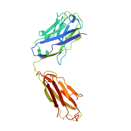
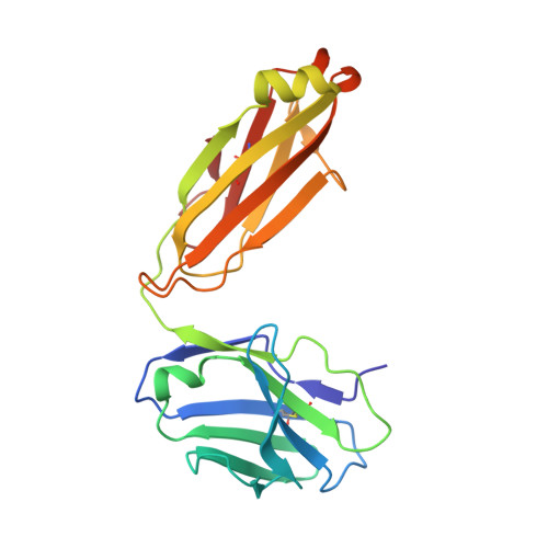
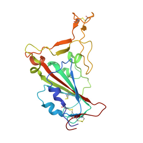
Etesevimab in combination with JS026 neutralizing SARS-CoV-2 and its variants.

Wang, F.Li, L.Dou, Y.Shi, R.Duan, X.Liu, H.Zhang, J.Liu, D.Wu, J.He, Y.Lan, J.Lu, B.Feng, H.Yan, J.

(2022) Emerg Microbes Infect 11: 548-551

  • DOI: https://doi.org/10.1080/22221751.2022.2032374
  • Primary Citation Related Structures: 
    7F7E

  • PubMed Abstract: 

    The neutralizing antibody is a potential therapeutic for the ongoing COVID-19 pandemic. As an antiviral agent, numerous mAbs recognize the epitopes that overlap with ACE2-binding sites in the SARS-CoV-2-RBD. Some studies have shown that residual changes on the spike protein can significantly decrease the efficiency of neutralizing antibodies. To address this issue, a therapeutic cocktail could be an effective countermeasure. In the present study, we isolated a fully human neutralizing antibody, JS026, from a convalescent patient. The comparative analysis revealed that JS026 binding to SARS-CoV-2-RBD mainly located between epitopes for class 2 and class 3 mAbs as opposed to that of class 1 (etesevimab) antibodies. A cocktail of etesevimab and JS026 increased neutralizing efficacy against both wild-type SARS-CoV-2 and the recent emergence of Alpha, Beta, Gamma, and Delta variants. JS026 and the cocktail reduced virus titers in the infected lungs of hACE2 transgenic mice and relieved pathological changes. These findings would benefit antibody-based therapeutic countermeasures in the treatment of COVID-19.


  • Organizational Affiliation
    • CAS Key Laboratory of Pathogenic Microbiology and Immunology, Institute of Microbiology, Chinese Academy of Sciences, Beijing, People's Republic of China.

Macromolecule Content 

  • Total Structure Weight: 68.67 kDa 
  • Atom Count: 4,835 
  • Modeled Residue Count: 630 
  • Deposited Residue Count: 630 
  • Unique protein chains: 3

Macromolecules

Find similar proteins by:|  3D Structure
Entity ID: 1
MoleculeChains  Sequence LengthOrganismDetailsImage
Heavy chain of A5-10 FabA [auth C]227Homo sapiensMutation(s): 0 
Find similar proteins by:|  3D Structure
Entity ID: 2
MoleculeChains  Sequence LengthOrganismDetailsImage
Light chain of A5-10 FabB [auth L]205Homo sapiensMutation(s): 0 
Find similar proteins by:|  3D Structure
Entity ID: 3
MoleculeChains  Sequence LengthOrganismDetailsImage
Spike protein S1C [auth E]198Severe acute respiratory syndrome coronavirus 2Mutation(s): 0 
Gene Names: S2
UniProt
Find proteins for P0DTC2 (Severe acute respiratory syndrome coronavirus 2)
Explore P0DTC2 
Go to UniProtKB:  P0DTC2
Entity Groups
Sequence Clusters30% Identity50% Identity70% Identity90% Identity95% Identity100% Identity
UniProt GroupP0DTC2
Glycosylation
Glycosylation Sites: 1Go to GlyGen: P0DTC2-1
Sequence Annotations
Expand
Reference Sequence

Small Molecules

Ligands 1 Unique
IDChains Name / Formula / InChI Key2D Diagram3D Interactions
NAG

Query on NAG


Download:Ideal Coordinates CCD File
D [auth E]2-acetamido-2-deoxy-beta-D-glucopyranose
C8 H15 N O6
OVRNDRQMDRJTHS-FMDGEEDCSA-N

Experimental Data & Validation

Experimental Data

  • Method: X-RAY DIFFRACTION
  • Resolution: 2.49 Å
  • R-Value Free:  0.256 (Depositor), 0.257 (DCC) 
  • R-Value Work:  0.214 (Depositor), 0.215 (DCC) 
  • R-Value Observed: 0.216 (Depositor) 
Space Group: H 3
Unit Cell:
Length ( Å )Angle ( ˚ )
a = 157.189α = 90
b = 157.189β = 90
c = 98.319γ = 120
Software Package:
Software NamePurpose
PHENIXrefinement
PDB_EXTRACTdata extraction
HKL-2000data reduction
HKL-2000data scaling
PHENIXphasing

Structure Validation

View Full Validation Report


Ligand Structure Quality Assessment 


Entry History 

Deposition Data

Revision History  (Full details and data files)

  • Version 1.0: 2022-02-02
    Type: Initial release
  • Version 1.1: 2022-02-23
    Changes: Database references
  • Version 1.2: 2023-11-29
    Changes: Data collection, Refinement description
  • Version 1.3: 2024-10-16
    Changes: Structure summary