7F69

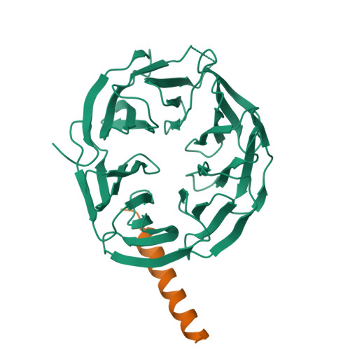
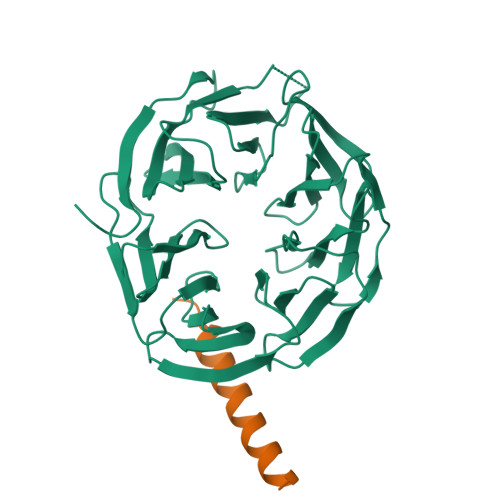
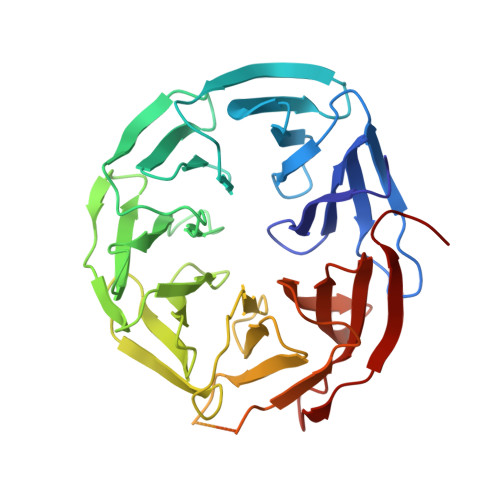
Crystal structure of WIPI2b in complex with ATG16L1


Experimental Data Snapshot

  • Method: X-RAY DIFFRACTION
  • Resolution: 1.50 Å
  • R-Value Free: 0.197 
  • R-Value Work: 0.182 
  • R-Value Observed: 0.183 

Starting Model: experimental
View more details

wwPDB Validation   3D Report Full Report


This is version 1.3 of the entry. See complete history


Literature

ATG16L1 adopts a dual-binding site mode to interact with WIPI2b in autophagy.

Gong, X.Wang, Y.Tang, Y.Wang, Y.Zhang, M.Li, M.Zhang, Y.Pan, L.

(2023) Sci Adv 9: eadf0824-eadf0824

  • DOI: https://doi.org/10.1126/sciadv.adf0824
  • Primary Citation of Related Structures:  
    7F69, 7XFR

  • PubMed Abstract: 

    Macroautophagy plays crucial roles in the regulation of cellular physiology and requires de novo synthesis of double-membrane autophagosomes, which relies on a specific interaction between autophagy-related 16L1 (ATG16L1) and WD repeat domain phosphoinositide-interacting protein 2b (WIPI2b). However, the molecular mechanism governing the interaction of ATG16L1 with WIPI2b remains elusive. Here, we find that ATG16L1 has two distinct binding sites for interacting with WIPI2b, the previously reported WIPI2b-binding site (WBS1) and the previously unidentified site (WBS2). We determine the crystal structures of WIPI2b with ATG16L1 WBS1 and WBS2, respectively, and elucidate the molecular mechanism underpinning the recruitment of ATG16L1 by WIPI2b. Moreover, we uncover that ATG16L1 WBS2 and its binding mode with WIPI2b is well conserved from yeast to mammals, unlike ATG16L1 WBS1. Last, our cell-based functional assays demonstrate that both ATG16L1 WBS1 and WBS2 are required for the effective autophagic flux. In conclusion, our findings provide mechanistic insights into the key ATG16L1/WIPI2b interaction in autophagy.


  • Organizational Affiliation

    State Key Laboratory of Bio-Organic and Natural Products Chemistry, Center for Excellence in Molecular Synthesis, Shanghai Institute of Organic Chemistry, University of Chinese Academy of Sciences, Chinese Academy of Sciences, Shanghai 200032, China.


Macromolecules
Find similar proteins by:  (by identity cutoff)  |  3D Structure
Entity ID: 1
MoleculeChains Sequence LengthOrganismDetailsImage
Isoform 2 of WD repeat domain phosphoinositide-interacting protein 2354Homo sapiensMutation(s): 0 
Gene Names: WIPI2CGI-50
UniProt & NIH Common Fund Data Resources
Find proteins for Q9Y4P8 (Homo sapiens)
Explore Q9Y4P8 
Go to UniProtKB:  Q9Y4P8
PHAROS:  Q9Y4P8
GTEx:  ENSG00000157954 
Entity Groups  
Sequence Clusters30% Identity50% Identity70% Identity90% Identity95% Identity100% Identity
UniProt GroupQ9Y4P8
Sequence Annotations
Expand
  • Reference Sequence
Find similar proteins by:  (by identity cutoff)  |  3D Structure
Entity ID: 2
MoleculeChains Sequence LengthOrganismDetailsImage
Isoform 2 of Autophagy-related protein 16-1B [auth C]34Homo sapiensMutation(s): 0 
Gene Names: ATG16L1APG16LUNQ9393/PRO34307
UniProt & NIH Common Fund Data Resources
Find proteins for Q676U5 (Homo sapiens)
Explore Q676U5 
Go to UniProtKB:  Q676U5
PHAROS:  Q676U5
GTEx:  ENSG00000085978 
Entity Groups  
Sequence Clusters30% Identity50% Identity70% Identity90% Identity95% Identity100% Identity
UniProt GroupQ676U5
Sequence Annotations
Expand
  • Reference Sequence
Experimental Data & Validation

Experimental Data

  • Method: X-RAY DIFFRACTION
  • Resolution: 1.50 Å
  • R-Value Free: 0.197 
  • R-Value Work: 0.182 
  • R-Value Observed: 0.183 
  • Space Group: P 63
Unit Cell:
Length ( Å )Angle ( ˚ )
a = 97.944α = 90
b = 97.944β = 90
c = 65.581γ = 120
Software Package:
Software NamePurpose
PHENIXrefinement
HKL-2000data reduction
HKL-2000data scaling
PHENIXphasing

Structure Validation

View Full Validation Report


Entry History & Funding Information

Deposition Data


Funding OrganizationLocationGrant Number
National Natural Science Foundation of China (NSFC)China21822705

Revision History  (Full details and data files)

  • Version 1.0: 2022-07-27
    Type: Initial release
  • Version 1.1: 2023-01-18
    Changes: Data collection
  • Version 1.2: 2023-03-08
    Changes: Database references
  • Version 1.3: 2023-11-29
    Changes: Data collection, Refinement description