7F3Q | pdb_00007f3q

SARS-CoV-2 RBD in complex with A5-10 Fab and A34-2 Fab


Experimental Data Snapshot

  • Method: ELECTRON MICROSCOPY
  • Resolution: 3.30 Å
  • Aggregation State: PARTICLE 
  • Reconstruction Method: SINGLE PARTICLE 

wwPDB Validation 3D Report Full Report

Validation slider image for 7F3Q

This is version 1.2 of the entry. See complete history

Literature

Development of neutralizing antibodies against SARS-CoV-2, using a high-throughput single-B-cell cloning method.

Dou, Y.Xu, K.Deng, Y.Q.Jia, Z.Lan, J.Xu, X.Zhang, G.Cao, T.Liu, P.Wang, X.Wang, X.Xu, L.Du, P.Qin, C.F.Liu, H.Li, Y.Wu, G.Wang, K.Lu, B.

(2023) Antib Ther 6: 76-86

  • DOI: https://doi.org/10.1093/abt/tbad002
  • Primary Citation Related Structures: 
    7F3Q

  • PubMed Abstract: 

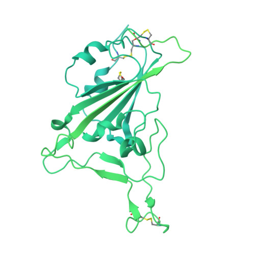
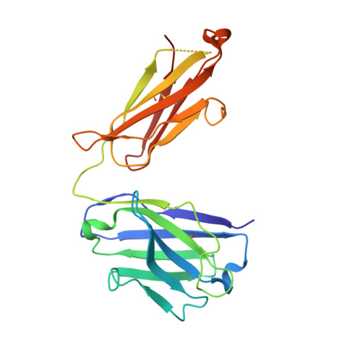
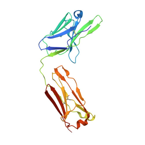
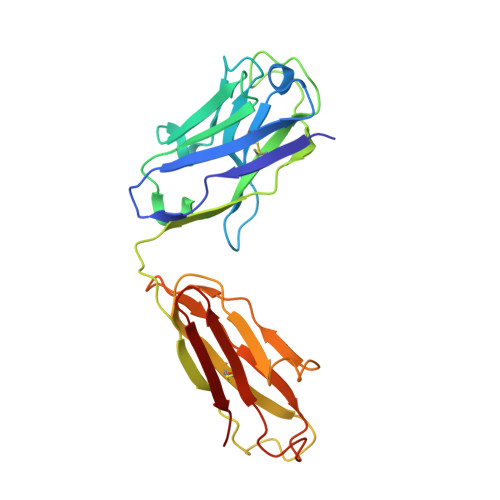
    Rapid and efficient strategies are needed to discover neutralizing antibodies (nAbs) from B cells derived from virus-infected patients. Here, we report a high-throughput single-B-cell cloning method for high-throughput isolation of nAbs targeting diverse epitopes on the SARS-CoV-2-RBD (receptor binding domain) from convalescent COVID-19 patients. This method is simple, fast and highly efficient in generating SARS-CoV-2-neutralizing antibodies from COVID-19 patients' B cells. Using this method, we have developed multiple nAbs against distinct SARS-CoV-2-RBD epitopes. CryoEM and crystallography revealed precisely how they bind RBD. In live virus assay, these nAbs are effective in blocking viral entry to the host cells. This simple and efficient method may be useful in developing human therapeutic antibodies for other diseases and next pandemic.


  • Organizational Affiliation
    • School of Pharmaceutical Sciences, IDG/McGovern Institute for Brain Research, Tsinghua University, Beijing 100084, China.

Macromolecule Content 

  • Total Structure Weight: 234.01 kDa 
  • Atom Count: 8,003 
  • Modeled Residue Count: 1,050 
  • Deposited Residue Count: 2,138 
  • Unique protein chains: 5

Macromolecules

Find similar proteins by:|  3D Structure
Entity ID: 1
MoleculeChains  Sequence LengthOrganismDetailsImage
Spike glycoprotein1,273Severe acute respiratory syndrome coronavirus 2Mutation(s): 0 
Gene Names: S2
UniProt
Find proteins for P0DTC2 (Severe acute respiratory syndrome coronavirus 2)
Explore P0DTC2 
Go to UniProtKB:  P0DTC2
Entity Groups
Sequence Clusters30% Identity50% Identity70% Identity90% Identity95% Identity100% Identity
UniProt GroupP0DTC2
Glycosylation
Glycosylation Sites: 1Go to GlyGen: P0DTC2-1
Sequence Annotations
Expand
Reference Sequence
Find similar proteins by:|  3D Structure
Entity ID: 2
MoleculeChains  Sequence LengthOrganismDetailsImage
Heavy chain of A34-2 FabB [auth H]219Homo sapiensMutation(s): 0 
Find similar proteins by:|  3D Structure
Entity ID: 3
MoleculeChains  Sequence LengthOrganismDetailsImage
Light chain of A34-2 FabC [auth L]214Homo sapiensMutation(s): 0 
Find similar proteins by:|  3D Structure
Entity ID: 4
MoleculeChains  Sequence LengthOrganismDetailsImage
Heavy chain of A5-10 FabD [auth C]227Homo sapiensMutation(s): 0 
Find similar proteins by:|  3D Structure
Entity ID: 5
MoleculeChains  Sequence LengthOrganismDetailsImage
Light chain of A5-10 FabE [auth B]205Homo sapiensMutation(s): 0 

Small Molecules

Experimental Data & Validation

Experimental Data

  • Method: ELECTRON MICROSCOPY
  • Resolution: 3.30 Å
  • Aggregation State: PARTICLE 
  • Reconstruction Method: SINGLE PARTICLE 

Structure Validation

View Full Validation Report


Entry History 

& Funding Information

Deposition Data


Funding OrganizationLocationGrant Number
Not funded--

Revision History  (Full details and data files)

  • Version 1.0: 2022-06-22
    Type: Initial release
  • Version 1.1: 2024-01-24
    Changes: Data collection, Database references
  • Version 1.2: 2024-10-30
    Changes: Data collection, Database references, Structure summary