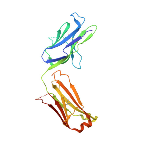
7F35

Crystal structure of anti S-gatifloxacin antibody Fab fragment in complex with S-gatifloxacin


Experimental Data Snapshot

  • Method: X-RAY DIFFRACTION
  • Resolution: 2.60 Å
  • R-Value Free: 0.235 
  • R-Value Work: 0.200 
  • R-Value Observed: 0.202 

Starting Model: experimental
View more details

wwPDB Validation   3D Report Full Report


Ligand Structure Quality Assessment 


This is version 1.1 of the entry. See complete history


Literature

Conformational adaptability determining antibody recognition to distomer: structure analysis of enantioselective antibody against chiral drug gatifloxacin

Wang, L.T.Xie, W.Jiao, W.Y.Zhang, C.Li, X.Xu, Z.Huang, X.Lei, H.T.Shen, X.

(2021) RSC Adv 11: 39534-39544


Macromolecules
Find similar proteins by:  (by identity cutoff)  |  3D Structure
Entity ID: 1
MoleculeChains Sequence LengthOrganismDetailsImage
Antibody Fab fragment light chain
A, C, E
218Mus musculusMutation(s): 0 
Entity Groups  
Sequence Clusters30% Identity50% Identity70% Identity90% Identity95% Identity100% Identity
Sequence Annotations
Expand
  • Reference Sequence
Find similar proteins by:  (by identity cutoff)  |  3D Structure
Entity ID: 2
MoleculeChains Sequence LengthOrganismDetailsImage
Antibody Fab fragment heavy chain
B, D, F
218Mus musculusMutation(s): 0 
Entity Groups  
Sequence Clusters30% Identity50% Identity70% Identity90% Identity95% Identity100% Identity
Sequence Annotations
Expand
  • Reference Sequence
Small Molecules
Ligands 7 Unique
IDChains Name / Formula / InChI Key2D Diagram3D Interactions
GFN (Subject of Investigation/LOI)
Query on GFN

Download Ideal Coordinates CCD File 
DA [auth E],
G [auth A],
Q [auth C]
1-cyclopropyl-6-fluoro-8-methoxy-7-[(3S)-3-methylpiperazin-1-yl]-4-oxo-1,4-dihydroquinoline-3-carboxylic acid
C19 H22 F N3 O4
XUBOMFCQGDBHNK-JTQLQIEISA-N
CIT
Query on CIT

Download Ideal Coordinates CCD File 
HA [auth F]CITRIC ACID
C6 H8 O7
KRKNYBCHXYNGOX-UHFFFAOYSA-N
SO4
Query on SO4

Download Ideal Coordinates CCD File 
EA [auth F]
FA [auth F]
GA [auth F]
K [auth B]
R [auth C]
EA [auth F],
FA [auth F],
GA [auth F],
K [auth B],
R [auth C],
S [auth C],
Z [auth D]
SULFATE ION
O4 S
QAOWNCQODCNURD-UHFFFAOYSA-L
GOL
Query on GOL

Download Ideal Coordinates CCD File 
T [auth C],
U [auth C]
GLYCEROL
C3 H8 O3
PEDCQBHIVMGVHV-UHFFFAOYSA-N
EDO
Query on EDO

Download Ideal Coordinates CCD File 
H [auth A]
I [auth A]
IA [auth F]
JA [auth F]
L [auth B]
H [auth A],
I [auth A],
IA [auth F],
JA [auth F],
L [auth B],
M [auth B],
N [auth B],
V [auth C]
1,2-ETHANEDIOL
C2 H6 O2
LYCAIKOWRPUZTN-UHFFFAOYSA-N
CL
Query on CL

Download Ideal Coordinates CCD File 
Y [auth C]CHLORIDE ION
Cl
VEXZGXHMUGYJMC-UHFFFAOYSA-M
NA
Query on NA

Download Ideal Coordinates CCD File 
AA [auth D]
BA [auth D]
CA [auth D]
J [auth A]
O [auth B]
AA [auth D],
BA [auth D],
CA [auth D],
J [auth A],
O [auth B],
P [auth B],
W [auth C],
X [auth C]
SODIUM ION
Na
FKNQFGJONOIPTF-UHFFFAOYSA-N
Experimental Data & Validation

Experimental Data

  • Method: X-RAY DIFFRACTION
  • Resolution: 2.60 Å
  • R-Value Free: 0.235 
  • R-Value Work: 0.200 
  • R-Value Observed: 0.202 
  • Space Group: P 42 21 2
Unit Cell:
Length ( Å )Angle ( ˚ )
a = 184.071α = 90
b = 184.071β = 90
c = 100.66γ = 90
Software Package:
Software NamePurpose
PHENIXrefinement
Aimlessdata scaling
PDB_EXTRACTdata extraction
XDSdata reduction
PHASERphasing

Structure Validation

View Full Validation Report


Ligand Structure Quality Assessment 


Entry History & Funding Information

Deposition Data


Funding OrganizationLocationGrant Number
National Basic Research Program of China (973 Program)China2017YFC1601700

Revision History  (Full details and data files)

  • Version 1.0: 2021-12-29
    Type: Initial release
  • Version 1.1: 2023-11-29
    Changes: Data collection, Refinement description