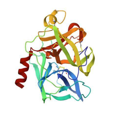
7EST

Interaction of the peptide CF3-LEU-ALA-NH-C6H4-CF3(TFLA) with porcine pancreatic elastase. X-ray studies at 1.8 Angstroms


Experimental Data Snapshot

  • Method: X-RAY DIFFRACTION
  • Resolution: 1.80 Å
  • R-Value Work: 0.190 

Starting Model: experimental
View more details

wwPDB Validation   3D Report Full Report


This is version 1.6 of the entry. See complete history


Literature

Interaction of the peptide CF3-Leu-Ala-NH-C6H4-CF3 (TFLA) with porcine pancreatic elastase. X-ray studies at 1.8 A.

de la Sierra, I.L.Papamichael, E.Sakarellos, C.Dimicoli, J.L.Prange, T.

(1990) J Mol Recognit 3: 36-44

  • DOI: https://doi.org/10.1002/jmr.300030104
  • Primary Citation of Related Structures:  
    6EST, 7EST

  • PubMed Abstract: 

    The peptide trifluoroacetyl-Leu-Ala-(p-trifluoromethylanilide), is a reversible inhibitor of pancreatic porcine elastase and is characterized by a Km of 2.5 x 10(-8) M. Co-crystals of the 1:1 complex were obtained in an acetate buffer + dimethylformamide solution at pH 5.7. Diffraction data were recorded on films at the LURE synchrotron facility. The inhibitor was localized on difference Fourier maps, and the refinement of the structure was performed by simulated annealing (XPLOR). The current agreement factor is R = 19% (for 13224 observed structure factors and 1.8 A effective resolution). The RMS deviations from ideality of bond distances and angles are 0.02 A and 2 degrees, respectively. The inhibitor molecule was found in the active site, bent around the side chain of Phe-215 in a geometry that resembles the previously reported structure of the CF3-Lys-Ala complex at 2.5 A, in a parallel beta-sheet association with the loop 214-216. The analysis of the close contacts (less than 3.5 A) indicates that the trifluoromethylamide bond interacts with the active site and not the Leu-Ala or Ala-anilide bonds. The two fluorinated groups of the inhibitor exhibit different specificities: the trifluoroacetyl group (N terminus) is tightly stacked between the two chain loops 191-195 and 213-215, while the trifluoromethylanilide (C terminus) shows less specificity and only a single contact.


  • Organizational Affiliation

    Université Paris-Sud, Orsay, France.


Macromolecules
Find similar proteins by:  (by identity cutoff)  |  3D Structure
Entity ID: 1
MoleculeChains Sequence LengthOrganismDetailsImage
ELASTASEA [auth E]240Sus scrofaMutation(s): 0 
EC: 3.4.21.36
UniProt
Find proteins for P00772 (Sus scrofa)
Explore P00772 
Go to UniProtKB:  P00772
Entity Groups  
Sequence Clusters30% Identity50% Identity70% Identity90% Identity95% Identity100% Identity
UniProt GroupP00772
Sequence Annotations
Expand
  • Reference Sequence
Small Molecules
Ligands 4 Unique
IDChains Name / Formula / InChI Key2D Diagram3D Interactions
0Z2
Query on 0Z2

Download Ideal Coordinates CCD File 
B [auth E]N-(trifluoroacetyl)-L-leucyl-N-[4-(trifluoromethyl)phenyl]-L-alaninamide
C18 H21 F6 N3 O3
COFHEZKAONGFAP-GWCFXTLKSA-N
SO4
Query on SO4

Download Ideal Coordinates CCD File 
C [auth E]SULFATE ION
O4 S
QAOWNCQODCNURD-UHFFFAOYSA-L
DMF
Query on DMF

Download Ideal Coordinates CCD File 
E,
F [auth E],
G [auth E],
H [auth E],
I [auth E]
DIMETHYLFORMAMIDE
C3 H7 N O
ZMXDDKWLCZADIW-UHFFFAOYSA-N
CA
Query on CA

Download Ideal Coordinates CCD File 
D [auth E]CALCIUM ION
Ca
BHPQYMZQTOCNFJ-UHFFFAOYSA-N
Biologically Interesting Molecules (External Reference) 1 Unique
Experimental Data & Validation

Experimental Data

  • Method: X-RAY DIFFRACTION
  • Resolution: 1.80 Å
  • R-Value Work: 0.190 
  • Space Group: P 21 21 21
Unit Cell:
Length ( Å )Angle ( ˚ )
a = 52.53α = 90
b = 57.47β = 90
c = 75.26γ = 90
Software Package:
Software NamePurpose
X-PLORrefinement

Structure Validation

View Full Validation Report


Entry History 

Deposition Data

Revision History  (Full details and data files)

  • Version 1.0: 1991-10-15
    Type: Initial release
  • Version 1.1: 2008-03-03
    Changes: Version format compliance
  • Version 1.2: 2011-07-13
    Changes: Atomic model, Database references, Derived calculations, Non-polymer description, Structure summary, Version format compliance
  • Version 1.3: 2011-12-21
    Changes: Database references
  • Version 1.4: 2012-12-12
    Changes: Other
  • Version 1.5: 2023-08-09
    Changes: Database references, Derived calculations, Other, Refinement description
  • Version 1.6: 2024-10-30
    Changes: Data collection, Structure summary