7ELL

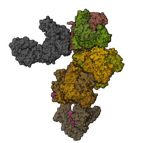
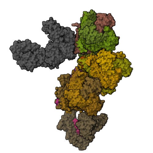
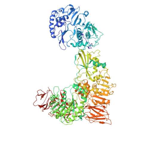
In situ structure of capping enzyme lambda2, penetration protein mu1 of mammalian reovirus capsid asymmetric unit.

  • Classification: VIRAL PROTEIN/TRANSFERASE
  • Organism(s): Mammalian orthoreovirus 3
  • Mutation(s): No 

  • Deposited: 2021-04-12 Released: 2021-10-20 
  • Deposition Author(s): Zhou, Z.H., Pan, M.
  • Funding Organization(s): National Institutes of Health/National Institute Of Allergy and Infectious Diseases (NIH/NIAID), National Science Foundation (NSF, United States)

Experimental Data Snapshot

  • Method: ELECTRON MICROSCOPY
  • Resolution: 3.80 Å
  • Aggregation State: PARTICLE 
  • Reconstruction Method: SINGLE PARTICLE 

wwPDB Validation   3D Report Full Report


This is version 1.1 of the entry. See complete history


Literature

Asymmetric reconstruction of mammalian reovirus reveals interactions among RNA, transcriptional factor mu2 and capsid proteins.

Pan, M.Alvarez-Cabrera, A.L.Kang, J.S.Wang, L.Fan, C.Zhou, Z.H.

(2021) Nat Commun 12: 4176-4176

  • DOI: https://doi.org/10.1038/s41467-021-24455-4
  • Primary Citation of Related Structures:  
    7ELH, 7ELL

  • PubMed Abstract: 

    Mammalian reovirus (MRV) is the prototypical member of genus Orthoreovirus of family Reoviridae. However, lacking high-resolution structures of its RNA polymerase cofactor μ2 and infectious particle, limits understanding of molecular interactions among proteins and RNA, and their contributions to virion assembly and RNA transcription. Here, we report the 3.3 Å-resolution asymmetric reconstruction of transcribing MRV and in situ atomic models of its capsid proteins, the asymmetrically attached RNA-dependent RNA polymerase (RdRp) λ3, and RdRp-bound nucleoside triphosphatase μ2 with a unique RNA-binding domain. We reveal molecular interactions among virion proteins and genomic and messenger RNA. Polymerase complexes in three Spinoreovirinae subfamily members are organized with different pseudo-D 3d symmetries to engage their highly diversified genomes. The above interactions and those between symmetry-mismatched receptor-binding σ1 trimers and RNA-capping λ2 pentamers balance competing needs of capsid assembly, external protein removal, and allosteric triggering of endogenous RNA transcription, before, during and after infection, respectively.


  • Organizational Affiliation

    CAS Key Laboratory of Interfacial Physics and Technology, Shanghai Institute of Applied Physics, Chinese Academy of Sciences, Shanghai, China.


Macromolecules
Find similar proteins by:  (by identity cutoff)  |  3D Structure
Entity ID: 1
MoleculeChains Sequence LengthOrganismDetailsImage
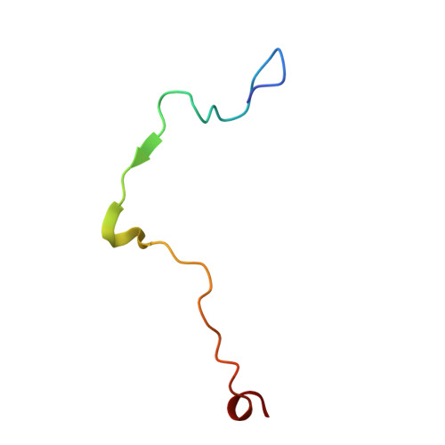
Mu141Mammalian orthoreovirus 3Mutation(s): 0 
UniProt
Find proteins for P11078 (Reovirus type 3 (strain Dearing))
Explore P11078 
Go to UniProtKB:  P11078
Entity Groups  
Sequence Clusters30% Identity50% Identity70% Identity90% Identity95% Identity100% Identity
UniProt GroupP11078
Sequence Annotations
Expand
  • Reference Sequence
Find similar proteins by:  (by identity cutoff)  |  3D Structure
Entity ID: 2
MoleculeChains Sequence LengthOrganismDetailsImage
Mu1666Mammalian orthoreovirus 3Mutation(s): 0 
UniProt
Find proteins for P11078 (Reovirus type 3 (strain Dearing))
Explore P11078 
Go to UniProtKB:  P11078
Entity Groups  
Sequence Clusters30% Identity50% Identity70% Identity90% Identity95% Identity100% Identity
UniProt GroupP11078
Sequence Annotations
Expand
  • Reference Sequence
Find similar proteins by:  (by identity cutoff)  |  3D Structure
Entity ID: 3
MoleculeChains Sequence LengthOrganismDetailsImage
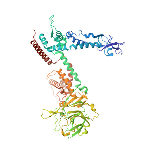
mRNA (guanine-N(7)-)-methyltransferaseU [auth K]1,289Mammalian orthoreovirus 3Mutation(s): 0 
EC: 2.1.1.56 (PDB Primary Data), 2.7.7.50 (PDB Primary Data)
UniProt
Find proteins for P11079 (Reovirus type 3 (strain Dearing))
Explore P11079 
Go to UniProtKB:  P11079
Entity Groups  
Sequence Clusters30% Identity50% Identity70% Identity90% Identity95% Identity100% Identity
UniProt GroupP11079
Sequence Annotations
Expand
  • Reference Sequence
Small Molecules
Ligands 1 Unique
IDChains Name / Formula / InChI Key2D Diagram3D Interactions
MYR (Subject of Investigation/LOI)
Query on MYR

Download Ideal Coordinates CCD File 
AA [auth f]
BA [auth g]
CA [auth h]
DA [auth i]
EA [auth j]
AA [auth f],
BA [auth g],
CA [auth h],
DA [auth i],
EA [auth j],
V [auth A],
W [auth b],
X [auth c],
Y [auth d],
Z [auth e]
MYRISTIC ACID
C14 H28 O2
TUNFSRHWOTWDNC-UHFFFAOYSA-N
Experimental Data & Validation

Experimental Data

  • Method: ELECTRON MICROSCOPY
  • Resolution: 3.80 Å
  • Aggregation State: PARTICLE 
  • Reconstruction Method: SINGLE PARTICLE 

Structure Validation

View Full Validation Report


Entry History & Funding Information

Deposition Data


Funding OrganizationLocationGrant Number
National Institutes of Health/National Institute Of Allergy and Infectious Diseases (NIH/NIAID)United StatesAI094386
National Institutes of Health/National Institute Of Allergy and Infectious Diseases (NIH/NIAID)United StatesGM071940
National Institutes of Health/National Institute Of Allergy and Infectious Diseases (NIH/NIAID)United StatesDE025567
National Institutes of Health/National Institute Of Allergy and Infectious Diseases (NIH/NIAID)United States1S10RR23057
National Institutes of Health/National Institute Of Allergy and Infectious Diseases (NIH/NIAID)United States1S10OD018111
National Institutes of Health/National Institute Of Allergy and Infectious Diseases (NIH/NIAID)United StatesU24GM116792
National Science Foundation (NSF, United States)United StatesDBI-1338135
National Science Foundation (NSF, United States)United StatesDMR-1548924

Revision History  (Full details and data files)

  • Version 1.0: 2021-10-20
    Type: Initial release
  • Version 1.1: 2024-11-06
    Changes: Data collection, Structure summary