7EFQ

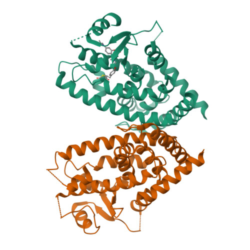
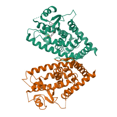
Crystal structure of hPPARgamma ligand binding domain complexed with rosiglitazone-based fluorescence probe


Experimental Data Snapshot

  • Method: X-RAY DIFFRACTION
  • Resolution: 2.30 Å
  • R-Value Free: 0.264 
  • R-Value Work: 0.224 

Starting Model: experimental
View more details

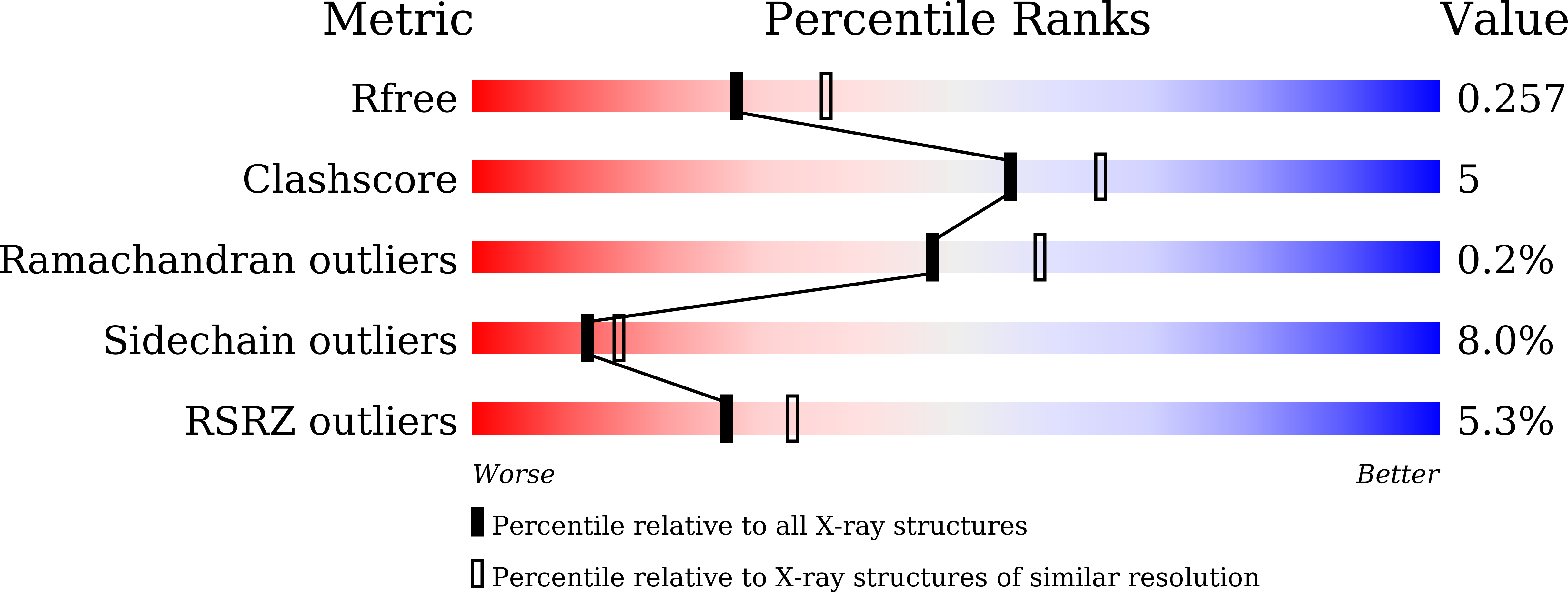
wwPDB Validation   3D Report Full Report


Ligand Structure Quality Assessment 


This is version 1.1 of the entry. See complete history


Literature

Synthesis of a Coumarin-Based PPAR gamma Fluorescence Probe for Competitive Binding Assay.

Yoshikawa, C.Ishida, H.Ohashi, N.Itoh, T.

(2021) Int J Mol Sci 22

  • DOI: https://doi.org/10.3390/ijms22084034
  • Primary Citation of Related Structures:  
    7EFQ

  • PubMed Abstract: 

    Peroxisome proliferator-activated receptor γ (PPARγ) is a molecular target of metabolic syndrome and inflammatory disease. PPARγ is an important nuclear receptor and numerous PPARγ ligands were developed to date; thus, efficient assay methods are important. Here, we investigated the incorporation of 7-diethylamino coumarin into the PPARγ agonist rosiglitazone and used the compound in a binding assay for PPARγ. PPARγ-ligand-incorporated 7-methoxycoumarin, 1 , showed weak fluorescence intensity in a previous report. We synthesized PPARγ-ligand-incorporating coumarin, 2 , in this report, and it enhanced the fluorescence intensity. The PPARγ ligand 2 maintained the rosiglitazone activity. The obtained partial agonist 6 appeared to act through a novel mechanism. The fluorescence intensity of 2 and 6 increased by binding to the ligand binding domain (LBD) of PPARγ and the affinity of reported PPARγ ligands were evaluated using the probe.


  • Organizational Affiliation

    Laboratory of Drug Design and Medicinal Chemistry, Showa Pharmaceutical University, 3-3165 Higashi-Tamagawagakuen, Machida, Tokyo 194-8543, Japan.


Macromolecules
Find similar proteins by:  (by identity cutoff)  |  3D Structure
Entity ID: 1
MoleculeChains Sequence LengthOrganismDetailsImage
Peroxisome proliferator-activated receptor gamma
A, B
276Homo sapiensMutation(s): 0 
Gene Names: PPARGNR1C3
UniProt & NIH Common Fund Data Resources
Find proteins for P37231 (Homo sapiens)
Explore P37231 
Go to UniProtKB:  P37231
PHAROS:  P37231
GTEx:  ENSG00000132170 
Entity Groups  
Sequence Clusters30% Identity50% Identity70% Identity90% Identity95% Identity100% Identity
UniProt GroupP37231
Sequence Annotations
Expand
  • Reference Sequence
Small Molecules
Ligands 1 Unique
IDChains Name / Formula / InChI Key2D Diagram3D Interactions
J3F (Subject of Investigation/LOI)
Query on J3F

Download Ideal Coordinates CCD File 
C [auth A](5S)-5-[[4-[2-[[7-(diethylamino)-2-oxidanylidene-chromen-4-yl]-methyl-amino]ethoxy]phenyl]methyl]-1,3-thiazolidine-2,4-dione
C26 H29 N3 O5 S
LTRSSWCLZRFXEC-QHCPKHFHSA-N
Experimental Data & Validation

Experimental Data

  • Method: X-RAY DIFFRACTION
  • Resolution: 2.30 Å
  • R-Value Free: 0.264 
  • R-Value Work: 0.224 
  • Space Group: C 1 2 1
Unit Cell:
Length ( Å )Angle ( ˚ )
a = 93.33α = 90
b = 61.9β = 102.29
c = 119.37γ = 90
Software Package:
Software NamePurpose
REFMACrefinement
Aimlessdata scaling
pointlessdata reduction
PHASERphasing
Cootmodel building

Structure Validation

View Full Validation Report


Ligand Structure Quality Assessment 


Entry History 

Deposition Data

Revision History  (Full details and data files)

  • Version 1.0: 2021-05-12
    Type: Initial release
  • Version 1.1: 2023-11-29
    Changes: Data collection, Database references, Derived calculations, Refinement description