7EC7

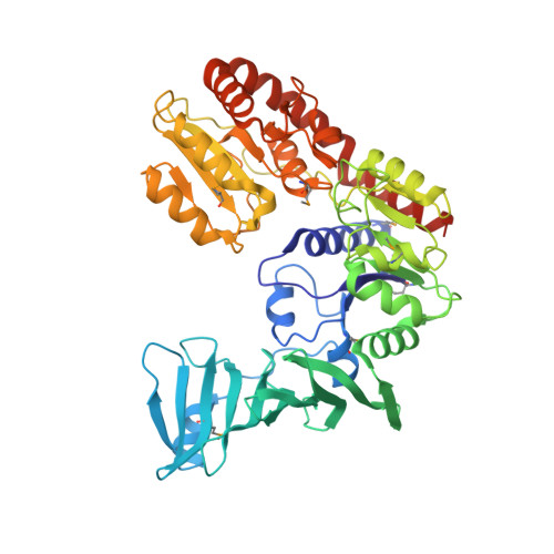
Crystal structure of SdgB (complexed with phosphate ions)


Experimental Data Snapshot

  • Method: X-RAY DIFFRACTION
  • Resolution: 3.20 Å
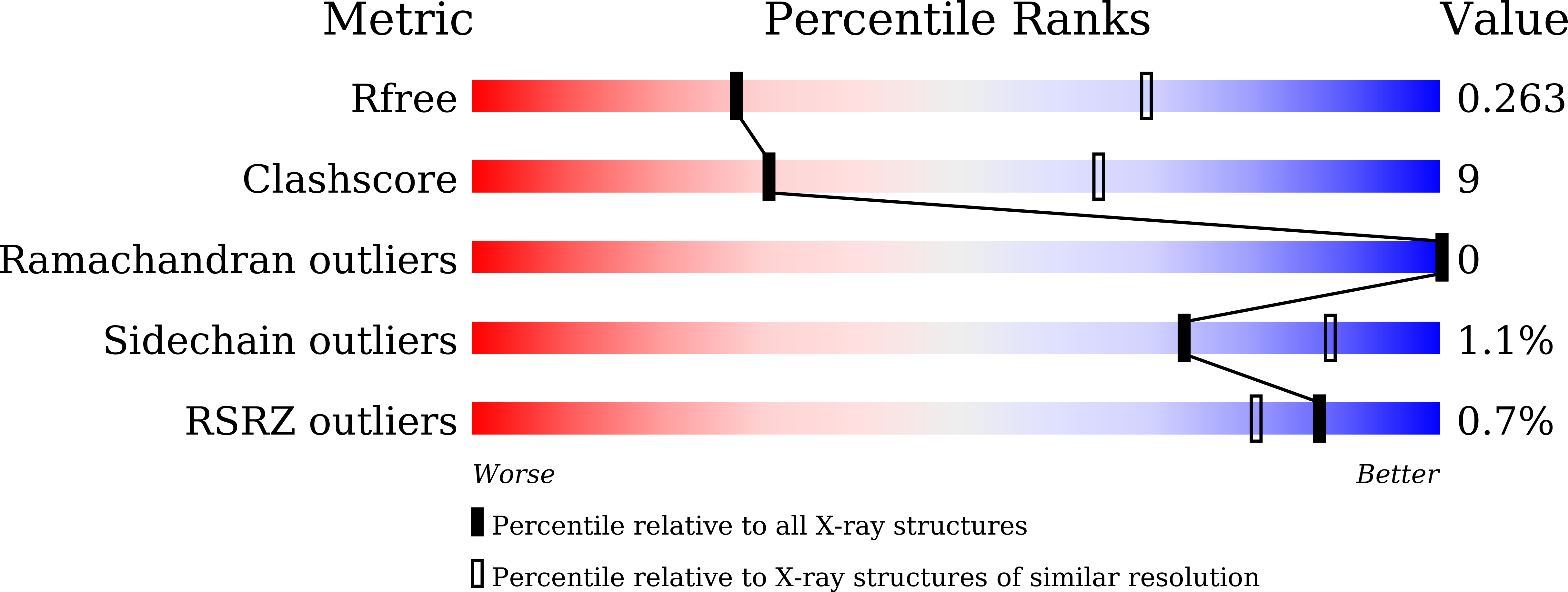
  • R-Value Free: 0.260 
  • R-Value Work: 0.196 
  • R-Value Observed: 0.199 

Starting Model: experimental
View more details

wwPDB Validation   3D Report Full Report


Ligand Structure Quality Assessment 


This is version 1.3 of the entry. See complete history


Literature

Structural basis for SdgB- and SdgA-mediated glycosylation of staphylococcal adhesive proteins.

Kim, D.G.Baek, I.Lee, Y.Kim, H.Kim, J.Y.Bang, G.Kim, S.Yoon, H.J.Han, B.W.Suh, S.W.Kim, H.S.

(2021) Acta Crystallogr D Struct Biol 77: 1460-1474

  • DOI: https://doi.org/10.1107/S2059798321010068
  • Primary Citation of Related Structures:  
    7EC1, 7EC3, 7EC6, 7EC7, 7VFK, 7VFL, 7VFM, 7VFN, 7VFO

  • PubMed Abstract: 

    The initiation of infection of host tissues by Staphylococcus aureus requires a family of staphylococcal adhesive proteins containing serine-aspartate repeat (SDR) domains, such as ClfA. The O-linked glycosylation of the long-chain SDR domain mediated by SdgB and SdgA is a key virulence factor that protects the adhesive SDR proteins against host proteolytic attack in order to promote successful tissue colonization, and has also been implicated in staphylococcal agglutination, which leads to sepsis and an immunodominant epitope for a strong antibody response. Despite the biological significance of these two glycosyltransferases involved in pathogenicity and avoidance of the host innate immune response, their structures and the molecular basis of their activity have not been investigated. This study reports the crystal structures of SdgB and SdgA from S. aureus as well as multiple structures of SdgB in complex with its substrates (for example UDP, N-acetylglucosamine or SDR peptides), products (glycosylated SDR peptides) or phosphate ions. Together with biophysical and biochemical analyses, this structural work uncovered the novel mechanism by which SdgB and SdgA carry out the glycosyl-transfer process to the long SDR region in SDR proteins. SdgB undergoes dynamic changes in its structure such as a transition from an open to a closed conformation upon ligand binding and takes diverse forms, both as a homodimer and as a heterodimer with SdgA. Overall, these findings not only elucidate the putative role of the three domains of SdgB in recognizing donor and acceptor substrates, but also provide new mechanistic insights into glycosylation of the SDR domain, which can serve as a starting point for the development of antibacterial drugs against staphylococcal infections.


  • Organizational Affiliation

    Research Institute, National Cancer Center, Goyang, Gyeonggi 10408, Republic of Korea.


Macromolecules
Find similar proteins by:  (by identity cutoff)  |  3D Structure
Entity ID: 1
MoleculeChains Sequence LengthOrganismDetailsImage
Glycosyl transferase, group 1 family protein
A, B
504Staphylococcus aureus subsp. aureus USA300Mutation(s): 0 
Gene Names: SAUSA300_0550
UniProt
Find proteins for A0A0H2XGN0 (Staphylococcus aureus (strain USA300))
Explore A0A0H2XGN0 
Go to UniProtKB:  A0A0H2XGN0
Entity Groups  
Sequence Clusters30% Identity50% Identity70% Identity90% Identity95% Identity100% Identity
UniProt GroupA0A0H2XGN0
Sequence Annotations
Expand
  • Reference Sequence
Small Molecules
Ligands 1 Unique
IDChains Name / Formula / InChI Key2D Diagram3D Interactions
PO4 (Subject of Investigation/LOI)
Query on PO4

Download Ideal Coordinates CCD File 
C [auth A]
D [auth A]
E [auth A]
F [auth A]
G [auth B]
C [auth A],
D [auth A],
E [auth A],
F [auth A],
G [auth B],
H [auth B],
I [auth B],
J [auth B],
K [auth B],
L [auth B],
M [auth B]
PHOSPHATE ION
O4 P
NBIIXXVUZAFLBC-UHFFFAOYSA-K
Modified Residues  1 Unique
IDChains TypeFormula2D DiagramParent
MSE
Query on MSE
A, B
L-PEPTIDE LINKINGC5 H11 N O2 SeMET
Experimental Data & Validation

Experimental Data

  • Method: X-RAY DIFFRACTION
  • Resolution: 3.20 Å
  • R-Value Free: 0.260 
  • R-Value Work: 0.196 
  • R-Value Observed: 0.199 
  • Space Group: P 32 2 1
Unit Cell:
Length ( Å )Angle ( ˚ )
a = 172.257α = 90
b = 172.257β = 90
c = 106.174γ = 120
Software Package:
Software NamePurpose
REFMACrefinement
PDB_EXTRACTdata extraction
HKL-2000data reduction
HKL-2000data scaling
MOLREPphasing

Structure Validation

View Full Validation Report


Ligand Structure Quality Assessment 


Entry History & Funding Information

Deposition Data


Funding OrganizationLocationGrant Number
National Research Foundation (NRF, Korea)Korea, Republic OfNRF-2020R1C1C1009512
National Research Foundation (NRF, Korea)Korea, Republic OfNRF-2017R1C1B2012225
National Research Foundation (NRF, Korea)Korea, Republic OfNRF-2015R1D1A4A01020265

Revision History  (Full details and data files)

  • Version 1.0: 2022-03-16
    Type: Initial release
  • Version 1.1: 2022-10-12
    Changes: Database references
  • Version 1.2: 2023-11-29
    Changes: Data collection, Refinement description
  • Version 1.3: 2024-10-30
    Changes: Structure summary