7EBD | pdb_00007ebd

Bacterial STING in complex with c-di-GMP


Experimental Data Snapshot

  • Method: X-RAY DIFFRACTION
  • Resolution: 2.25 Å
  • R-Value Free: 
    0.249 (Depositor), 0.250 (DCC) 
  • R-Value Work: 
    0.188 (Depositor), 0.190 (DCC) 
  • R-Value Observed: 
    0.191 (Depositor) 

wwPDB Validation 3D Report Full Report

Validation slider image for 7EBD

Ligand Structure Quality Assessment 


This is version 1.2 of the entry. See complete history

Literature

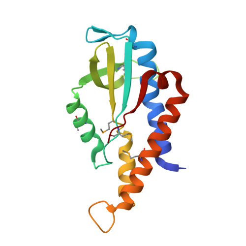
Crystal structure and functional implication of bacterial STING.

Ko, T.-P.Wang, Y.-C.Yang, C.-S.Hou, M.-H.Chen, C.-J.Chiu, Y.-F.Chen, Y.

(2022) Nat Commun 13: 26-26

  • DOI: https://doi.org/10.1038/s41467-021-26583-3
  • Primary Citation Related Structures: 
    7EBD, 7EBL

  • PubMed Abstract: 

    Mammalian innate immune sensor STING (STimulator of INterferon Gene) was recently found to originate from bacteria. During phage infection, bacterial STING sense c-di-GMP generated by the CD-NTase (cGAS/DncV-like nucleotidyltransferase) encoded in the same operon and signal suicide commitment as a defense strategy that restricts phage propagation. However, the precise binding mode of c-di-GMP to bacterial STING and the specific recognition mechanism are still elusive. Here, we determine two complex crystal structures of bacterial STING/c-di-GMP, which provide a clear picture of how c-di-GMP is distinguished from other cyclic dinucleotides. The protein-protein interactions further reveal the driving force behind filament formation of bacterial STING. Finally, we group the bacterial STING into two classes based on the conserved motif in β-strand lid, which dictate their ligand specificity and oligomerization mechanism, and propose an evolution-based model that describes the transition from c-di-GMP-dependent signaling in bacteria to 2'3'-cGAMP-dependent signaling in eukaryotes.


  • Organizational Affiliation
    • Institute of Biological Chemistry, Academia Sinica, Taipei, 115, Taiwan.

Macromolecule Content 

  • Total Structure Weight: 38.07 kDa 
  • Atom Count: 2,845 
  • Modeled Residue Count: 316 
  • Deposited Residue Count: 322 
  • Unique protein chains: 1

Macromolecules

Find similar proteins by:|  3D Structure
Entity ID: 1
MoleculeChains  Sequence LengthOrganismDetailsImage
TIR-like domain-containing protein
A, B
161Prevotella corporisMutation(s): 0 
Gene Names: HMPREF3226_02659
EC: 3.2.2.5
UniProt
Find proteins for A0A133PTK7 (Prevotella corporis)
Explore A0A133PTK7 
Go to UniProtKB:  A0A133PTK7
Entity Groups
Sequence Clusters30% Identity50% Identity70% Identity90% Identity95% Identity100% Identity
UniProt GroupA0A133PTK7
Sequence Annotations
Expand
Reference Sequence

Small Molecules

Ligands 2 Unique
IDChains Name / Formula / InChI Key2D Diagram3D Interactions
C2E
(Subject of Investigation/LOI)

Query on C2E


Download:Ideal Coordinates CCD File
E [auth B]9,9'-[(2R,3R,3aS,5S,7aR,9R,10R,10aS,12S,14aR)-3,5,10,12-tetrahydroxy-5,12-dioxidooctahydro-2H,7H-difuro[3,2-d:3',2'-j][1,3,7,9,2,8]tetraoxadiphosphacyclododecine-2,9-diyl]bis(2-amino-1,9-dihydro-6H-purin-6-one)
C20 H24 N10 O14 P2
PKFDLKSEZWEFGL-MHARETSRSA-N
ACT

Query on ACT


Download:Ideal Coordinates CCD File
C [auth A],
D [auth B]
ACETATE ION
C2 H3 O2
QTBSBXVTEAMEQO-UHFFFAOYSA-M
Modified Residues  1 Unique
IDChains TypeFormula2D DiagramParent
MSE
Query on MSE
A, B
L-PEPTIDE LINKINGC5 H11 N O2 SeMET

Experimental Data & Validation

Experimental Data

  • Method: X-RAY DIFFRACTION
  • Resolution: 2.25 Å
  • R-Value Free:  0.249 (Depositor), 0.250 (DCC) 
  • R-Value Work:  0.188 (Depositor), 0.190 (DCC) 
  • R-Value Observed: 0.191 (Depositor) 
Space Group: P 21 21 2
Unit Cell:
Length ( Å )Angle ( ˚ )
a = 94.534α = 90
b = 86.995β = 90
c = 35.223γ = 90
Software Package:
Software NamePurpose
PHENIXrefinement
HKL-2000data scaling
PHENIXphasing

Structure Validation

View Full Validation Report


Ligand Structure Quality Assessment 


Entry History 

& Funding Information

Deposition Data


Funding OrganizationLocationGrant Number
Academia Sinica (Taiwan)Taiwan--

Revision History  (Full details and data files)

  • Version 1.0: 2021-09-15
    Type: Initial release
  • Version 1.1: 2022-01-19
    Changes: Database references
  • Version 1.2: 2024-10-16
    Changes: Data collection, Refinement description, Structure summary