7E4R | pdb_00007e4r

Crystal structure of tubulin in complex with D-DM1-SMe


Experimental Data Snapshot

  • Method: X-RAY DIFFRACTION
  • Resolution: 2.60 Å
  • R-Value Free: 
    0.265 (Depositor), 0.271 (DCC) 
  • R-Value Work: 
    0.224 (Depositor), 0.229 (DCC) 
  • R-Value Observed: 
    0.225 (Depositor) 

Starting Model: experimental
View more details

wwPDB Validation 3D Report Full Report

Validation slider image for 7E4R

Ligand Structure Quality Assessment 


This is version 1.1 of the entry. See complete history

Literature

C3 ester side chain plays a pivotal role in the antitumor activity of Maytansinoids.

Li, W.Huang, M.Li, Y.Xia, A.Tan, L.Zhang, Z.Wang, Y.Yang, J.

(2021) Biochem Biophys Res Commun 566: 197-203

  • DOI: https://doi.org/10.1016/j.bbrc.2021.05.071
  • Primary Citation Related Structures: 
    7E4Q, 7E4R, 7E4Z

  • PubMed Abstract: 

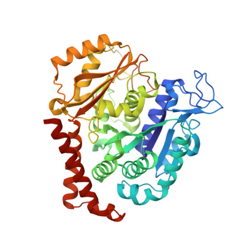
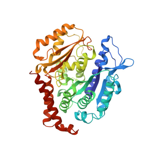
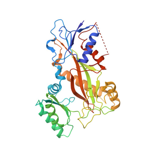
    Maytansinoids, the chemical derivatives of Maytansine, are commonly used as potent cytotoxic payloads in antibody-drug conjugates (ADC). Structure-activity-relationship studies had identified the C3 ester side chain as a critical element for antitumor activity of maytansinoids. The maytansinoids bearing the methyl group at C3 position with D configuration were about 100 to 400-fold less cytotoxic than their corresponding L-epimers toward various cell lines. The detailed mechanism of how chirality affects the anticancer activity remains elusive. In this study, we determined the high-resolution crystal structure of tubulin in complex with maytansinol, L-DM1-SMe and D-DM1-SMe. And we found the carbonyl oxygen atom of the ester moiety and the tail thiomethyl group at C3 side chain of L-DM1-SMe form strong intramolecular interaction with the hydroxyl at position 9 and the benzene ring, respectively, fixing the bioactive conformation and enhancing the binding affinity. Additionally, ligand-based and structure-based virtually screening methods were used to screen the commercially macrocyclic compounds library, and 15 macrocyclic structures were picketed out as putatively new maytansine-site inhibitors. Our study provides a possible strategy for the rational discovery of next-generation maytansine site inhibitors.


  • Organizational Affiliation
    • State Key Laboratory of Biotherapy and Collaborative Innovation Center for Biotherapy, West China Hospital, Sichuan University, Chengdu, 610041, PR China. Electronic address: liwentingo@qq.com.

Macromolecule Content 

  • Total Structure Weight: 258.5 kDa 
  • Atom Count: 17,814 
  • Modeled Residue Count: 2,180 
  • Deposited Residue Count: 2,260 
  • Unique protein chains: 4

Macromolecules

Find similar proteins by:|  3D Structure
Entity ID: 1
MoleculeChains  Sequence LengthOrganismDetailsImage
Tubulin alpha-1B chain
A, C
440Bos taurusMutation(s): 0 
EC: 3.6.5
UniProt
Find proteins for P81947 (Bos taurus)
Explore P81947 
Go to UniProtKB:  P81947
Entity Groups
Sequence Clusters30% Identity50% Identity70% Identity90% Identity95% Identity100% Identity
UniProt GroupP81947
Sequence Annotations
Expand
Reference Sequence
Find similar proteins by:|  3D Structure
Entity ID: 2
MoleculeChains  Sequence LengthOrganismDetailsImage
Tubulin beta-2B chain
B, D
431Bos taurusMutation(s): 0 
Gene Names: TUBB2B
UniProt
Find proteins for Q6B856 (Bos taurus)
Explore Q6B856 
Go to UniProtKB:  Q6B856
Entity Groups
Sequence Clusters30% Identity50% Identity70% Identity90% Identity95% Identity100% Identity
UniProt GroupQ6B856
Sequence Annotations
Expand
Reference Sequence
Find similar proteins by:|  3D Structure
Entity ID: 3
MoleculeChains  Sequence LengthOrganismDetailsImage
Stathmin-4138Rattus norvegicusMutation(s): 0 
Gene Names: Stmn4
UniProt
Find proteins for P63043 (Rattus norvegicus)
Explore P63043 
Go to UniProtKB:  P63043
Entity Groups
Sequence Clusters30% Identity50% Identity70% Identity90% Identity95% Identity100% Identity
UniProt GroupP63043
Sequence Annotations
Expand
Reference Sequence
Find similar proteins by:|  3D Structure
Entity ID: 4
MoleculeChains  Sequence LengthOrganismDetailsImage
Tubulin tyrosine ligase380Gallus gallusMutation(s): 0 
Gene Names: TTL

Small Molecules

Ligands 7 Unique
IDChains Name / Formula / InChI Key2D Diagram3D Interactions
HZ0

Query on HZ0


Download:Ideal Coordinates CCD File
Q [auth D](10E,12E)-86-chloro-14-hydroxy-85,14-dimethoxy-33,2,7,10-tetramethyl-12,6-dioxo-7-aza-1(6,4)-oxazinana-3(2,3)-oxirana-8(1,3)-benzenacyclotetradecaphane-10,12-dien-4-yl N-methyl-N-(3-(methylsulfinothioyl)propanoyl)-D-alaninate
C36 H50 Cl N3 O10 S2
ZLUUPZXOPGORNG-YXIMMCLRSA-N
GTP

Query on GTP


Download:Ideal Coordinates CCD File
G [auth A],
M [auth C],
P [auth D]
GUANOSINE-5'-TRIPHOSPHATE
C10 H16 N5 O14 P3
XKMLYUALXHKNFT-UUOKFMHZSA-N
ACP

Query on ACP


Download:Ideal Coordinates CCD File
S [auth F]PHOSPHOMETHYLPHOSPHONIC ACID ADENYLATE ESTER
C11 H18 N5 O12 P3
UFZTZBNSLXELAL-IOSLPCCCSA-N
GDP

Query on GDP


Download:Ideal Coordinates CCD File
J [auth B]GUANOSINE-5'-DIPHOSPHATE
C10 H15 N5 O11 P2
QGWNDRXFNXRZMB-UUOKFMHZSA-N
MES

Query on MES


Download:Ideal Coordinates CCD File
L [auth B]2-(N-MORPHOLINO)-ETHANESULFONIC ACID
C6 H13 N O4 S
SXGZJKUKBWWHRA-UHFFFAOYSA-N
CA

Query on CA


Download:Ideal Coordinates CCD File
I [auth A],
O [auth C]
CALCIUM ION
Ca
BHPQYMZQTOCNFJ-UHFFFAOYSA-N
MG

Query on MG


Download:Ideal Coordinates CCD File
H [auth A],
K [auth B],
N [auth C],
R [auth D]
MAGNESIUM ION
Mg
JLVVSXFLKOJNIY-UHFFFAOYSA-N

Experimental Data & Validation

Experimental Data

  • Method: X-RAY DIFFRACTION
  • Resolution: 2.60 Å
  • R-Value Free:  0.265 (Depositor), 0.271 (DCC) 
  • R-Value Work:  0.224 (Depositor), 0.229 (DCC) 
  • R-Value Observed: 0.225 (Depositor) 
Space Group: P 21 21 21
Unit Cell:
Length ( Å )Angle ( ˚ )
a = 104.982α = 90
b = 157.229β = 90
c = 182.574γ = 90
Software Package:
Software NamePurpose
HKL-3000data scaling
PHENIXrefinement
PDB_EXTRACTdata extraction
HKL-3000data reduction
PHASERphasing

Structure Validation

View Full Validation Report


Ligand Structure Quality Assessment 


Entry History 

& Funding Information

Deposition Data


Funding OrganizationLocationGrant Number
National Natural Science Foundation of China (NSFC)China81703553

Revision History  (Full details and data files)

  • Version 1.0: 2021-07-07
    Type: Initial release
  • Version 1.1: 2023-11-29
    Changes: Data collection, Database references, Refinement description