7E2O | pdb_00007e2o

X-ray Crystal structure of PPARgamma R288H mutant.


Experimental Data Snapshot

  • Method: X-RAY DIFFRACTION
  • Resolution: 3.20 Å
  • R-Value Free: 
    0.313 (Depositor), 0.310 (DCC) 
  • R-Value Work: 
    0.234 (Depositor), 0.235 (DCC) 
  • R-Value Observed: 
    0.238 (Depositor) 

Starting Model: experimental
View more details

wwPDB Validation 3D Report Full Report

Validation slider image for 7E2O

This is version 1.1 of the entry. See complete history

Literature

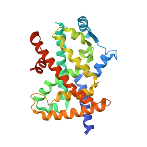
Structural Insights into the Loss-of-Function R288H Mutant of Human PPAR gamma.

Egawa, D.Ogiso, T.Nishikata, K.Yamamoto, K.Itoh, T.

(2021) Biol Pharm Bull 44: 1196-1201

  • DOI: https://doi.org/10.1248/bpb.b21-00253
  • Primary Citation Related Structures: 
    7E2O

  • PubMed Abstract: 

    Peroxisome proliferator-activated receptor gamma (PPARγ) is a nuclear receptor and the molecular target of thiazolidinedione-class antidiabetic drugs. It has been reported that the loss of function R288H mutation in the human PPARγ ligand-binding domain (LBD) may be associated with the onset of colon cancer. A previous in vitro study showed that this mutation dampens 15-deoxy-Δ 12,14 -prostaglandin J2 (15d-PGJ2, a natural PPARγ agonist)-dependent transcriptional activation; however, it is poorly understood why the function of the R288H mutant is impaired and what role this arginine (Arg) residue plays. In this study, we found that the apo-form of R288H PPARγ mutant displays several altered conformational arrangements of the amino acid side chains in LBD: 1) the loss of a salt bridge between Arg288 and Glu295 leads to increased helix 3 movement; 2) closer proximity of Gln286 and His449 via a hydrogen bond, and closer proximity of Cys285 and Phe363 via hydrophobic interaction, stabilize the helix 3-helix 11 interaction; and 3) there is steric hindrance between Cys285/Gln286/Ser289/His449 and the flexible ligands 15d-PGJ2, 6-oxotetracosahexaenoic acid (6-oxoTHA), and 17-oxodocosahexaenoic acid (17-oxoDHA). These results suggest why Arg288 plays an important role in ligand binding and why the R288H mutation is disadvantageous for flexible ligand binding.


  • Organizational Affiliation
    • Laboratory of Drug Design and Medicinal Chemistry, Showa Pharmaceutical University.

Macromolecule Content 

  • Total Structure Weight: 62.86 kDa 
  • Atom Count: 3,935 
  • Modeled Residue Count: 500 
  • Deposited Residue Count: 552 
  • Unique protein chains: 1

Macromolecules

Find similar proteins by:|  3D Structure
Entity ID: 1
MoleculeChains  Sequence LengthOrganismDetailsImage
Peroxisome proliferator-activated receptor gamma
A, B
276Homo sapiensMutation(s): 1 
Gene Names: PPARGNR1C3
UniProt & NIH Common Fund Data Resources
Find proteins for P37231 (Homo sapiens)
Explore P37231 
Go to UniProtKB:  P37231
PHAROS:  P37231
GTEx:  ENSG00000132170 
Entity Groups
Sequence Clusters30% Identity50% Identity70% Identity90% Identity95% Identity100% Identity
UniProt GroupP37231
Sequence Annotations
Expand
Reference Sequence

Experimental Data & Validation

Experimental Data

  • Method: X-RAY DIFFRACTION
  • Resolution: 3.20 Å
  • R-Value Free:  0.313 (Depositor), 0.310 (DCC) 
  • R-Value Work:  0.234 (Depositor), 0.235 (DCC) 
  • R-Value Observed: 0.238 (Depositor) 
Space Group: C 1 2 1
Unit Cell:
Length ( Å )Angle ( ˚ )
a = 91.04α = 90
b = 60.92β = 102.24
c = 116.73γ = 90
Software Package:
Software NamePurpose
REFMACrefinement
PDB_EXTRACTdata extraction
iMOSFLMdata reduction
Aimlessdata scaling
REFMACphasing

Structure Validation

View Full Validation Report


Entry History 

Deposition Data

Revision History  (Full details and data files)

  • Version 1.0: 2022-01-19
    Type: Initial release
  • Version 1.1: 2023-11-29
    Changes: Data collection, Refinement description