7DY7 | pdb_00007dy7

Discovery of Novel Small-molecule Inhibitors of PD-1/PD-L1 Axis that Promotes PD-L1 Internalization and Degradation


Experimental Data Snapshot

  • Method: X-RAY DIFFRACTION
  • Resolution: 2.42 Å
  • R-Value Free: 
    0.240 (Depositor), 0.246 (DCC) 
  • R-Value Work: 
    0.196 (Depositor), 0.201 (DCC) 
  • R-Value Observed: 
    0.198 (Depositor) 

Starting Model: experimental
View more details

wwPDB Validation 3D Report Full Report

Validation slider image for 7DY7

Ligand Structure Quality Assessment 


This is version 1.3 of the entry. See complete history

Literature

Discovery of Small-Molecule Inhibitors of the PD-1/PD-L1 Axis That Promote PD-L1 Internalization and Degradation.

Wang, T.Cai, S.Cheng, Y.Zhang, W.Wang, M.Sun, H.Guo, B.Li, Z.Xiao, Y.Jiang, S.

(2022) J Med Chem 65: 3879-3893

  • DOI: https://doi.org/10.1021/acs.jmedchem.1c01682
  • Primary Citation Related Structures: 
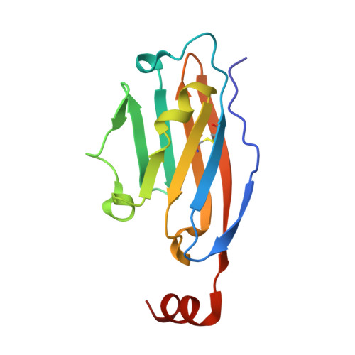
    7DY7

  • PubMed Abstract: 

    Several monoclonal antibodies targeting the programmed cell death-1/programmed cell death-ligand 1 (PD-1/PD-L1) pathway have been used successfully in anticancer immunotherapy. Inherent limitations of antibody-based therapies remain, however, and alternative small-molecule inhibitors that can block the PD-1/PD-L1 axis are urgent needed. Herein, we report the discovery of compound 17 as a bifunctional inhibitor of PD-1/PD-L1 interactions. 17 inhibits PD-1/PD-L1 interactions and promotes dimerization, internalization, and degradation of PD-L1. 17 promotes cell-surface PD-L1 internalized into the cytosol and induces the degradation of PD-L1 in tumor cells through a lysosome-dependent pathway. Furthermore, 17 suppresses tumor growth in vivo by activating antitumor immunity. These results demonstrate that 17 targets the PD-1/PD-L1 axis and induces PD-L1 degradation.


  • Organizational Affiliation
    • State Key Laboratory of Natural Medicines and Department of Medicinal Chemistry, School of Pharmacy, China Pharmaceutical University, Nanjing 210009, China.

Macromolecule Content 

  • Total Structure Weight: 29.21 kDa 
  • Atom Count: 2,058 
  • Modeled Residue Count: 252 
  • Deposited Residue Count: 252 
  • Unique protein chains: 1

Macromolecules

Find similar proteins by:|  3D Structure
Entity ID: 1
MoleculeChains  Sequence LengthOrganismDetailsImage
Programmed cell death 1 ligand 1A [auth B],
B [auth A]
126Homo sapiensMutation(s): 0 
Gene Names: CD274B7H1PDCD1L1PDCD1LG1PDL1
UniProt & NIH Common Fund Data Resources
Find proteins for Q9NZQ7 (Homo sapiens)
Explore Q9NZQ7 
Go to UniProtKB:  Q9NZQ7
PHAROS:  Q9NZQ7
GTEx:  ENSG00000120217 
Entity Groups
Sequence Clusters30% Identity50% Identity70% Identity90% Identity95% Identity100% Identity
UniProt GroupQ9NZQ7
Sequence Annotations
Expand
Reference Sequence

Small Molecules

Ligands 1 Unique
IDChains Name / Formula / InChI Key2D Diagram3D Interactions
HOU
(Subject of Investigation/LOI)

Query on HOU


Download:Ideal Coordinates CCD File
C [auth B]2-[[3-[[5-(2-methyl-3-phenyl-phenyl)-1,3,4-oxadiazol-2-yl]amino]phenyl]methylamino]ethanol
C24 H24 N4 O2
YBHXMLDVPTWSAS-UHFFFAOYSA-N

Experimental Data & Validation

Experimental Data

  • Method: X-RAY DIFFRACTION
  • Resolution: 2.42 Å
  • R-Value Free:  0.240 (Depositor), 0.246 (DCC) 
  • R-Value Work:  0.196 (Depositor), 0.201 (DCC) 
  • R-Value Observed: 0.198 (Depositor) 
Space Group: C 1 2 1
Unit Cell:
Length ( Å )Angle ( ˚ )
a = 134.57α = 90
b = 32.682β = 103.96
c = 73.603γ = 90
Software Package:
Software NamePurpose
PHENIXrefinement
PDB_EXTRACTdata extraction
HKL-3000data reduction
HKL-2000data scaling
MOLREPphasing

Structure Validation

View Full Validation Report


Ligand Structure Quality Assessment 


Entry History 

Deposition Data

Revision History  (Full details and data files)

  • Version 1.0: 2022-01-26
    Type: Initial release
  • Version 1.1: 2022-04-27
    Changes: Database references
  • Version 1.2: 2023-11-29
    Changes: Data collection, Refinement description
  • Version 1.3: 2024-10-16
    Changes: Structure summary