7DWR | pdb_00007dwr

Structure of Sulfolobus solfataricus SegA-ADP complex bound to DNA


Experimental Data Snapshot

  • Method: X-RAY DIFFRACTION
  • Resolution: 2.80 Å
  • R-Value Free: 
    0.298 (Depositor), 0.294 (DCC) 
  • R-Value Work: 
    0.239 (Depositor), 0.238 (DCC) 
  • R-Value Observed: 
    0.243 (Depositor) 

Starting Model: experimental
View more details

wwPDB Validation 3D Report Full Report

Validation slider image for 7DWR

Ligand Structure Quality Assessment 


This is version 1.2 of the entry. See complete history

Literature

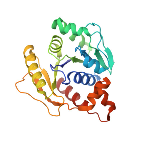
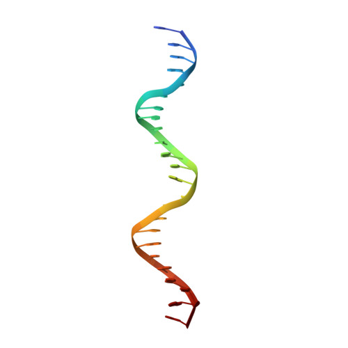
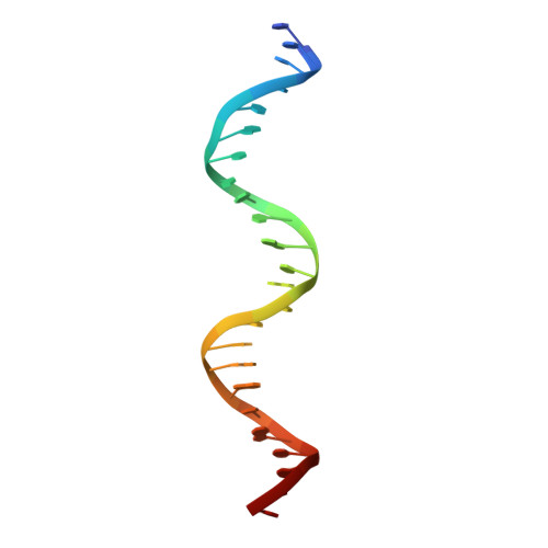
Chromosome segregation in Archaea: SegA- and SegB-DNA complex structures provide insights into segrosome assembly.

Yen, C.Y.Lin, M.G.Chen, B.W.Ng, I.W.Read, N.Kabli, A.F.Wu, C.T.Shen, Y.Y.Chen, C.H.Barilla, D.Sun, Y.J.Hsiao, C.D.

(2021) Nucleic Acids Res 49: 13150-13164

  • DOI: https://doi.org/10.1093/nar/gkab1155
  • Primary Citation Related Structures: 
    7DUT, 7DUV, 7DV2, 7DV3, 7DWR

  • PubMed Abstract: 

    Genome segregation is a vital process in all organisms. Chromosome partitioning remains obscure in Archaea, the third domain of life. Here, we investigated the SegAB system from Sulfolobus solfataricus. SegA is a ParA Walker-type ATPase and SegB is a site-specific DNA-binding protein. We determined the structures of both proteins and those of SegA-DNA and SegB-DNA complexes. The SegA structure revealed an atypical, novel non-sandwich dimer that binds DNA either in the presence or in the absence of ATP. The SegB structure disclosed a ribbon-helix-helix motif through which the protein binds DNA site specifically. The association of multiple interacting SegB dimers with the DNA results in a higher order chromatin-like structure. The unstructured SegB N-terminus plays an essential catalytic role in stimulating SegA ATPase activity and an architectural regulatory role in segrosome (SegA-SegB-DNA) formation. Electron microscopy results also provide a compact ring-like segrosome structure related to chromosome organization. These findings contribute a novel mechanistic perspective on archaeal chromosome segregation.


  • Organizational Affiliation
    • Institute of Bioinformatics and Structural Biology, National Tsing Hua University, Hsinchu 300, Taiwan.

Macromolecule Content 

  • Total Structure Weight: 128.58 kDa 
  • Atom Count: 8,947 
  • Modeled Residue Count: 976 
  • Deposited Residue Count: 976 
  • Unique protein chains: 1
  • Unique nucleic acid chains: 2

Macromolecules


Find similar proteins by:|  3D Structure
Entity ID: 1
MoleculeChains  Sequence LengthOrganismDetailsImage
SOJ protein (Soj)
A, B, C, D
220Saccharolobus solfataricus P2Mutation(s): 0 
Gene Names: sojSSO0034
UniProt
Find proteins for Q981B3 (Saccharolobus solfataricus (strain ATCC 35092 / DSM 1617 / JCM 11322 / P2))
Explore Q981B3 
Go to UniProtKB:  Q981B3
Entity Groups
Sequence Clusters30% Identity50% Identity70% Identity90% Identity95% Identity100% Identity
UniProt GroupQ981B3
Sequence Annotations
Expand
Reference Sequence
Find similar nucleic acids by:  Sequence
Entity ID: 2
MoleculeChains LengthOrganismImage
DNA (5'-D(P*AP*GP*GP*GP*TP*GP*TP*TP*CP*CP*AP*CP*GP*TP*GP*AP*AP*AP*CP*AP*GP*GP*GP*A)-3')
E, G
24synthetic construct
Sequence Annotations
Expand
Reference Sequence
Find similar nucleic acids by:  Sequence
Entity ID: 3
MoleculeChains LengthOrganismImage
DNA (5'-D(P*TP*CP*CP*CP*TP*GP*TP*TP*TP*CP*AP*CP*GP*TP*GP*GP*AP*AP*CP*AP*CP*CP*CP*T)-3')
F, H
24synthetic construct
Sequence Annotations
Expand
Reference Sequence

Small Molecules

Ligands 2 Unique
IDChains Name / Formula / InChI Key2D Diagram3D Interactions
ADP
(Subject of Investigation/LOI)

Query on ADP


Download:Ideal Coordinates CCD File
I [auth A],
K [auth B],
M [auth C],
O [auth D]
ADENOSINE-5'-DIPHOSPHATE
C10 H15 N5 O10 P2
XTWYTFMLZFPYCI-KQYNXXCUSA-N
MG

Query on MG


Download:Ideal Coordinates CCD File
J [auth A],
L [auth B],
N [auth C],
P [auth D]
MAGNESIUM ION
Mg
JLVVSXFLKOJNIY-UHFFFAOYSA-N

Experimental Data & Validation

Experimental Data

  • Method: X-RAY DIFFRACTION
  • Resolution: 2.80 Å
  • R-Value Free:  0.298 (Depositor), 0.294 (DCC) 
  • R-Value Work:  0.239 (Depositor), 0.238 (DCC) 
  • R-Value Observed: 0.243 (Depositor) 
Space Group: C 1 2 1
Unit Cell:
Length ( Å )Angle ( ˚ )
a = 220α = 90
b = 60.541β = 127.01
c = 132.143γ = 90
Software Package:
Software NamePurpose
PHENIXrefinement
SCALEPACKdata scaling
PDB_EXTRACTdata extraction
HKL-2000data reduction
PHASERphasing

Structure Validation

View Full Validation Report


Ligand Structure Quality Assessment 


Entry History 

& Funding Information

Deposition Data


Funding OrganizationLocationGrant Number
Ministry of Science and Technology (MoST, Taiwan)Taiwan--

Revision History  (Full details and data files)

  • Version 1.0: 2021-12-22
    Type: Initial release
  • Version 1.1: 2022-02-16
    Changes: Database references
  • Version 1.2: 2023-11-29
    Changes: Data collection, Refinement description