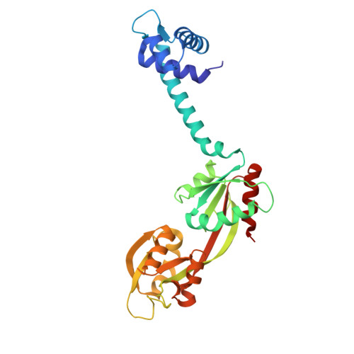
7DWN

Crystal structure of Vibrio fischeri DarR in complex with DNA reveals the transcriptional activation mechanism of LTTR family members


Experimental Data Snapshot

  • Method: X-RAY DIFFRACTION
  • Resolution: 2.32 Å
  • R-Value Free: 0.275 
  • R-Value Work: 0.228 
  • R-Value Observed: 0.230 

Starting Model: experimental
View more details

wwPDB Validation   3D Report Full Report


Ligand Structure Quality Assessment 


This is version 1.1 of the entry. See complete history


Literature

Crystal structure details of Vibrio fischeri DarR and mutant DarR-M202I from LTTR family reveals their activation mechanism.

Wang, W.Wu, H.Xiao, Q.Zhou, H.Li, M.Xu, Q.Wang, Q.Yu, F.He, J.

(2021) Int J Biol Macromol 183: 2354-2363

  • DOI: https://doi.org/10.1016/j.ijbiomac.2021.05.186
  • Primary Citation of Related Structures:  
    7DWN, 7DWO

  • PubMed Abstract: 

    DarR, a novel member of the LTTR family derived from Vibrio fischeri, activates transcription in response to d-Asp and regulates the overexpression of the racD genes encoding a putative aspartate racemase, RacD. Here, the crystal structure of full-length DarR and its mutant DarR-M202I were obtained by X-ray crystallography. According to the electron density map analysis of full-length DarR, the effector binding site of DarR is occupied by 2-Morpholinoethanesulfonic acid monohydrate (MES), which could interact with amino acids in the effector binding site and stabilize the effector binding site. Furthermore, we elaborated the structure of DarR-M202I, where methionine is replaced by isoleucine resulting in overexpression of the downstream operon. By comparing DarR-MES and DarR-M202I, we found similar behavior of DarR-MES in terms of the stability of the RD active pocket and the deflection angle of the DBD. The Isothermal titration calorimetry and Gel-filtration chromatography experiments showed that only when the target DNA sequence of a particular quasi-palindromic sequence exceeds 19 bp, DarR can effectively bind to racD promoter. This study will help enhance our understanding of the mechanism in the transcriptional regulation of LTTR family transcription factors.


  • Organizational Affiliation

    Shanghai Institute of Applied Physics, Chinese Academy of Sciences, Shanghai 201800, China; University of Chinese Academy of Sciences, Beijing 100049, China.


Macromolecules
Find similar proteins by:  (by identity cutoff)  |  3D Structure
Entity ID: 1
MoleculeChains Sequence LengthOrganismDetailsImage
Predicted DNA-binding transcriptional regulator
A, B, C, D
293Aliivibrio fischeri ES114Mutation(s): 0 
Gene Names: yjiEVF_1545
UniProt
Find proteins for Q5E4K6 (Aliivibrio fischeri (strain ATCC 700601 / ES114))
Explore Q5E4K6 
Go to UniProtKB:  Q5E4K6
Entity Groups  
Sequence Clusters30% Identity50% Identity70% Identity90% Identity95% Identity100% Identity
UniProt GroupQ5E4K6
Sequence Annotations
Expand
  • Reference Sequence
Experimental Data & Validation

Experimental Data

  • Method: X-RAY DIFFRACTION
  • Resolution: 2.32 Å
  • R-Value Free: 0.275 
  • R-Value Work: 0.228 
  • R-Value Observed: 0.230 
  • Space Group: P 1
Unit Cell:
Length ( Å )Angle ( ˚ )
a = 50.688α = 74.46
b = 77.456β = 78.16
c = 86.797γ = 89.86
Software Package:
Software NamePurpose
PHENIXrefinement
Aimlessdata scaling
PDB_EXTRACTdata extraction
XDSdata reduction
PHASERphasing

Structure Validation

View Full Validation Report


Ligand Structure Quality Assessment 


Entry History & Funding Information

Deposition Data


Funding OrganizationLocationGrant Number
National Natural Science Foundation of China (NSFC)China31971121
National Natural Science Foundation of China (NSFC)China81861138047
National Natural Science Foundation of China (NSFC)ChinaU1932130

Revision History  (Full details and data files)

  • Version 1.0: 2021-07-07
    Type: Initial release
  • Version 1.1: 2023-11-29
    Changes: Data collection, Database references, Refinement description