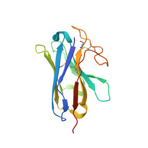
7DTY

Structural basis of ligand selectivity conferred by the human glucose-dependent insulinotropic polypeptide receptor


Experimental Data Snapshot

  • Method: ELECTRON MICROSCOPY
  • Resolution: 2.98 Å
  • Aggregation State: PARTICLE 
  • Reconstruction Method: SINGLE PARTICLE 

wwPDB Validation   3D Report Full Report


This is version 1.2 of the entry. See complete history


Literature

Structural insights into hormone recognition by the human glucose-dependent insulinotropic polypeptide receptor.

Zhao, F.Zhang, C.Zhou, Q.Hang, K.Zou, X.Chen, Y.Wu, F.Rao, Q.Dai, A.Yin, W.Shen, D.D.Zhang, Y.Xia, T.Stevens, R.C.Xu, H.E.Yang, D.Zhao, L.Wang, M.W.

(2021) Elife 10

  • DOI: https://doi.org/10.7554/eLife.68719
  • Primary Citation of Related Structures:  
    7DTY

  • PubMed Abstract: 

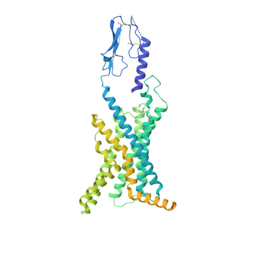
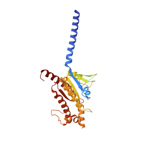
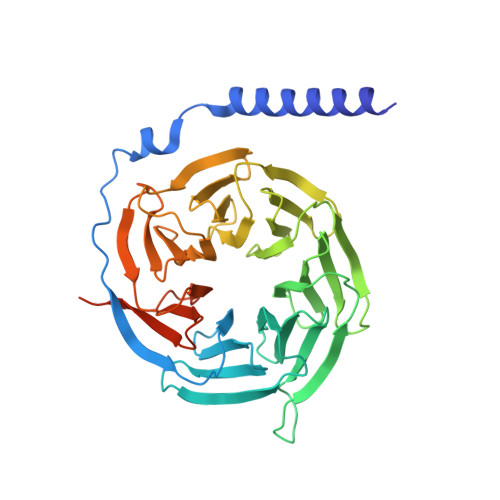
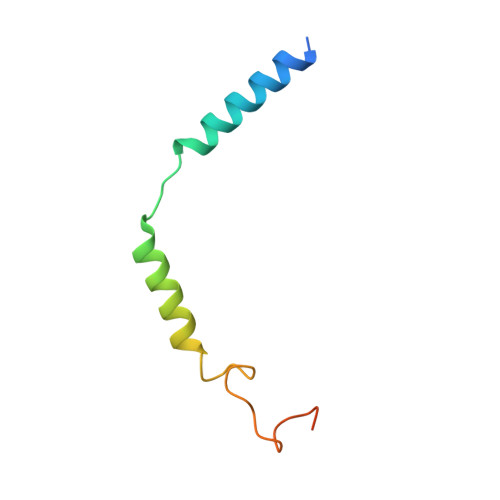
    Glucose-dependent insulinotropic polypeptide (GIP) is a peptide hormone that exerts crucial metabolic functions by binding and activating its cognate receptor, GIPR. As an important therapeutic target, GIPR has been subjected to intensive structural studies without success. Here, we report the cryo-EM structure of the human GIPR in complex with GIP and a G s heterotrimer at a global resolution of 2.9 Å. GIP adopts a single straight helix with its N terminus dipped into the receptor transmembrane domain (TMD), while the C terminus is closely associated with the extracellular domain and extracellular loop 1. GIPR employs conserved residues in the lower half of the TMD pocket to recognize the common segments shared by GIP homologous peptides, while uses non-conserved residues in the upper half of the TMD pocket to interact with residues specific for GIP. These results provide a structural framework of hormone recognition and GIPR activation.


  • Organizational Affiliation

    School of Pharmacy, Fudan University, Shanghai, China.


Macromolecules
Find similar proteins by:  (by identity cutoff)  |  3D Structure
Entity ID: 1
MoleculeChains Sequence LengthOrganismDetailsImage
human glucose-dependent insulinotropic polypeptide receptorA [auth R]573Homo sapiensMutation(s): 0 
UniProt & NIH Common Fund Data Resources
Find proteins for P48546 (Homo sapiens)
Go to UniProtKB:  P48546
PHAROS:  P48546
GTEx:  ENSG00000010310 
Sequence Annotations
Expand
  • Reference Sequence
Find similar proteins by:  (by identity cutoff)  |  3D Structure
Entity ID: 2
MoleculeChains Sequence LengthOrganismDetailsImage
Gastric inhibitory polypeptideB [auth P]42Homo sapiensMutation(s): 0 
UniProt & NIH Common Fund Data Resources
Find proteins for P09681 (Homo sapiens)
Go to UniProtKB:  P09681
PHAROS:  P09681
GTEx:  ENSG00000159224 
Sequence Annotations
Expand
  • Reference Sequence
Find similar proteins by:  (by identity cutoff)  |  3D Structure
Entity ID: 3
MoleculeChains Sequence LengthOrganismDetailsImage
Guanine nucleotide-binding protein G(s) subunit alpha isoforms shortC [auth A]394Bos taurusMutation(s): 0 
Gene Names: GNASGNAS1
UniProt
Find proteins for P04896 (Bos taurus)
Go to UniProtKB:  P04896
Sequence Annotations
Expand
  • Reference Sequence
Find similar proteins by:  (by identity cutoff)  |  3D Structure
Entity ID: 4
MoleculeChains Sequence LengthOrganismDetailsImage
Guanine nucleotide-binding protein G(I)/G(S)/G(T) subunit beta-1D [auth B]371Rattus norvegicusMutation(s): 0 
Gene Names: Gnb1
UniProt
Find proteins for P54311 (Rattus norvegicus)
Go to UniProtKB:  P54311
Sequence Annotations
Expand
  • Reference Sequence
Find similar proteins by:  (by identity cutoff)  |  3D Structure
Entity ID: 5
MoleculeChains Sequence LengthOrganismDetailsImage
Guanine nucleotide-binding protein G(I)/G(S)/G(O) subunit gamma-2E [auth G]71Bos taurusMutation(s): 0 
Gene Names: GNG2
UniProt
Find proteins for P63212 (Bos taurus)
Go to UniProtKB:  P63212
Sequence Annotations
Expand
  • Reference Sequence
Find similar proteins by:  (by identity cutoff)  |  3D Structure
Entity ID: 6
MoleculeChains Sequence LengthOrganismDetailsImage
Nanobody-35F [auth N]140synthetic constructMutation(s): 0 
Sequence Annotations
Expand
  • Reference Sequence
Experimental Data & Validation

Experimental Data

  • Method: ELECTRON MICROSCOPY
  • Resolution: 2.98 Å
  • Aggregation State: PARTICLE 
  • Reconstruction Method: SINGLE PARTICLE 

Structure Validation

View Full Validation Report


Entry History & Funding Information

Deposition Data


Funding OrganizationLocationGrant Number
National Natural Science Foundation of China (NSFC)China81872915
National Natural Science Foundation of China (NSFC)China21704064
National Natural Science Foundation of China (NSFC)China81773792
National Natural Science Foundation of China (NSFC)China32071203

Revision History  (Full details and data files)

  • Version 1.0: 2021-08-04
    Type: Initial release
  • Version 1.1: 2021-08-11
    Changes: Database references
  • Version 1.2: 2024-10-16
    Changes: Data collection, Structure summary