7DR4 | pdb_00007dr4

Complex of anti-human IL-2 antibody and human IL-2


Experimental Data Snapshot

  • Method: X-RAY DIFFRACTION
  • Resolution: 2.49 Å
  • R-Value Free: 
    0.258 (Depositor), 0.257 (DCC) 
  • R-Value Work: 
    0.195 (Depositor), 0.192 (DCC) 
  • R-Value Observed: 
    0.196 (Depositor) 

Starting Model: experimental
View more details

wwPDB Validation 3D Report Full Report

Validation slider image for 7DR4

This is version 1.2 of the entry. See complete history

Literature

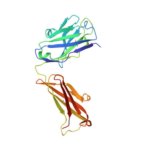
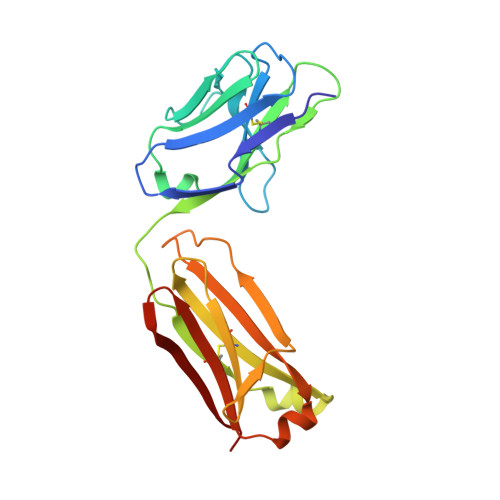
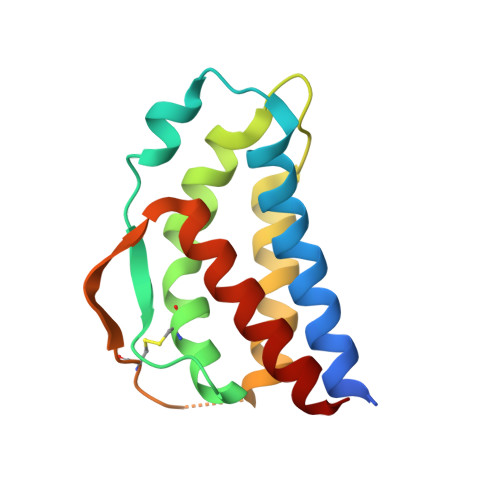
Crystal structure of human interleukin-2 in complex with TCB2, a new antibody-drug candidate with antitumor activity.

Kim, J.Lee, J.Y.Park, S.Y.Lee, Y.J.Kim, M.S.

(2021) Oncoimmunology 10: 1899671-1899671

  • DOI: https://doi.org/10.1080/2162402X.2021.1899671
  • Primary Citation Related Structures: 
    7DR4

  • PubMed Abstract: 

    Immunotherapy via interleukin-2 (IL-2) mediated activation of anti-tumor immune response is a promising approach for cancer treatment. The multi-potent cytokine, IL-2 has a central role in immune cell activation and homeostasis. Since IL-2 preferentially activates immunosuppressive T regulatory cells by IL-2Rα dependent manner, blocking IL-2:IL-2Rα interaction is a key to amplify the IL-2 activity in effector T cells toward anti-tumor response. Anti-IL-2 monoclonal antibodies are good candidates to control the IL-2:IL-2Rα interaction. In a previous study, we developed a new IL-2Rα mimetic antibody, TCB2, and showed that the human IL-2(hIL-2):TCB2 complex can stimulate T effector cells specifically and elicit potent anti-cancer immunotherapeutic effect, especially when administered in combination with immune checkpoint inhibitors. To understand the molecular mechanism, we determined the crystal structure of TCB2-Fab in a complex with hIL-2 at 2.5 Å resolution. Our structural analysis reveals that TCB2 binds to the central area of the hIL-2Rα binding region on hIL-2, and binding angle and epitope are different from previously known hIL-2Rα mimicking antibody NARA1 which recognizes the top part of hIL-2. TCB2 binding to hIL-2 also induces an allosteric effect that increases the affinity for the hetero-dimeric hIL-2 receptor, IL-2R(β + γ), on effector T cells.


  • Organizational Affiliation
    • Department of Life Sciences, Pohang University of Science and Technology (POSTECH), Pohang, Gyeongbuk, Republic of Korea.

Macromolecule Content 

  • Total Structure Weight: 252.25 kDa 
  • Atom Count: 17,347 
  • Modeled Residue Count: 2,174 
  • Deposited Residue Count: 2,284 
  • Unique protein chains: 3

Macromolecules

Find similar proteins by:|  3D Structure
Entity ID: 1
MoleculeChains  Sequence LengthOrganismDetailsImage
anti-human IL-2 antibody, mouse Ig G, heavy chainA [auth H],
C [auth A],
E [auth C],
G [auth E]
224Mus musculusMutation(s): 0 
Find similar proteins by:|  3D Structure
Entity ID: 2
MoleculeChains  Sequence LengthOrganismDetailsImage
anti-human IL-2 antibody, mouse Ig G, kappa chainB [auth L],
D [auth B],
F [auth D],
H [auth F]
214Mus musculusMutation(s): 0 
Find similar proteins by:|  3D Structure
Entity ID: 3
MoleculeChains  Sequence LengthOrganismDetailsImage
Interleukin-2I [auth G],
J [auth I],
K [auth J],
L [auth K]
133Homo sapiensMutation(s): 0 
Gene Names: IL2
UniProt & NIH Common Fund Data Resources
Find proteins for P60568 (Homo sapiens)
Explore P60568 
Go to UniProtKB:  P60568
PHAROS:  P60568
GTEx:  ENSG00000109471 
Entity Groups
Sequence Clusters30% Identity50% Identity70% Identity90% Identity95% Identity100% Identity
UniProt GroupP60568
Sequence Annotations
Expand
Reference Sequence

Experimental Data & Validation

Experimental Data

  • Method: X-RAY DIFFRACTION
  • Resolution: 2.49 Å
  • R-Value Free:  0.258 (Depositor), 0.257 (DCC) 
  • R-Value Work:  0.195 (Depositor), 0.192 (DCC) 
  • R-Value Observed: 0.196 (Depositor) 
Space Group: P 1 21 1
Unit Cell:
Length ( Å )Angle ( ˚ )
a = 79.334α = 90
b = 72.261β = 92.711
c = 210.93γ = 90
Software Package:
Software NamePurpose
PHENIXrefinement
HKL-2000data reduction
HKL-2000data scaling
PHENIXphasing

Structure Validation

View Full Validation Report


Entry History 

Deposition Data

Revision History  (Full details and data files)

  • Version 1.0: 2021-04-14
    Type: Initial release
  • Version 1.1: 2023-11-29
    Changes: Data collection, Database references, Refinement description
  • Version 1.2: 2024-11-06
    Changes: Structure summary