7DN9

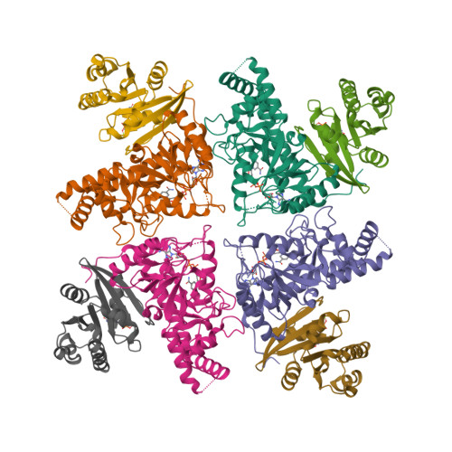
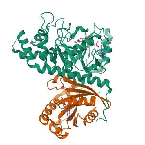
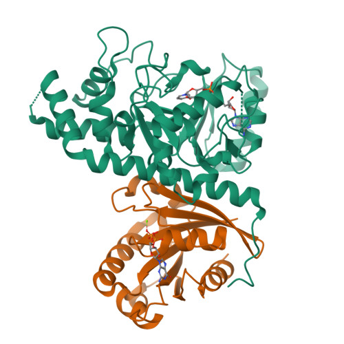
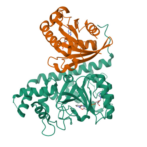
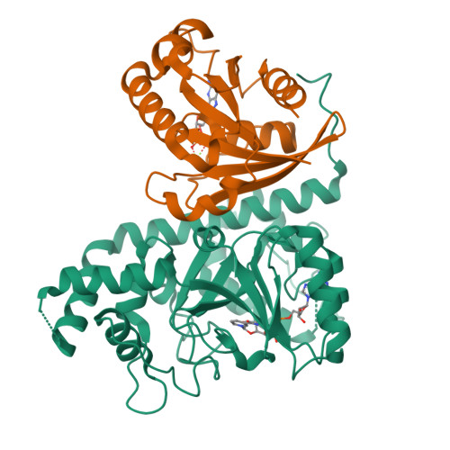
Crystal structure of Salmonella effector in complex with NAD and host co-factor ARF1


Experimental Data Snapshot

  • Method: X-RAY DIFFRACTION
  • Resolution: 3.29 Å
  • R-Value Free: 
    0.277 (Depositor), 0.280 (DCC) 
  • R-Value Work: 
    0.236 (Depositor), 0.240 (DCC) 
  • R-Value Observed: 
    0.239 (Depositor) 

wwPDB Validation   3D Report Full Report


Ligand Structure Quality Assessment 

Created with Raphaël 2.3.0Worse 01 BetterLigand structure goodness of fit to experimental dataBest fitted NADClick on this verticalbar to view details

This is version 1.2 of the entry. See complete history


Literature

ARF GTPases activate Salmonella effector SopF to ADP-ribosylate host V-ATPase and inhibit endomembrane damage-induced autophagy.

Xu, Y.Cheng, S.Zeng, H.Zhou, P.Ma, Y.Li, L.Liu, X.Shao, F.Ding, J.

(2022) Nat Struct Mol Biol 29: 67-77

  • DOI: https://doi.org/10.1038/s41594-021-00710-6
  • Primary Citation of Related Structures:  
    7DN8, 7DN9

  • PubMed Abstract: 

    Selective autophagy helps eukaryotes to cope with endogenous dangers or foreign invaders; its initiation often involves membrane damage. By studying a Salmonella effector SopF, we recently identified the vacuolar ATPase (V-ATPase)-ATG16L1 axis that initiates bacteria-induced autophagy. Here we show that SopF is an ADP-ribosyltransferase specifically modifying Gln124 of ATP6V0C in V-ATPase. We identify GTP-bound ADP-ribosylation factor (ARF) GTPases as a cofactor required for SopF functioning. Crystal structures of SopF-ARF1 complexes not only reveal structural basis of SopF ADP-ribosyltransferase activity but also a unique effector-binding mode adopted by ARF GTPases. Further, the N terminus of ARF1, although dispensable for high-affinity binding to SopF, is critical for activating SopF to modify ATP6V0C. Moreover, lysosome or Golgi damage-induced autophagic LC3 activation is inhibited by SopF or Q124A mutation of ATP6V0C, thus also mediated by the V-ATPase-ATG16L1 axis. In this process, the V-ATPase functions to sense membrane damages, which can be uncoupled from its proton-pumping activity.


  • Organizational Affiliation

    National Institute of Biological Sciences, Beijing, China.


Macromolecules
Find similar proteins by:  (by identity cutoff)  |  3D Structure
Entity ID: 1
MoleculeChains Sequence LengthOrganismDetailsImage
Putative cytoplasmic protein
A, C, E, G
312Salmonella enterica subsp. enterica serovar Typhimurium str. LT2Mutation(s): 1 
Gene Names: STM1239
UniProt
Find proteins for Q8ZPY9 (Salmonella typhimurium (strain LT2 / SGSC1412 / ATCC 700720))
Explore Q8ZPY9 
Go to UniProtKB:  Q8ZPY9
Entity Groups  
Sequence Clusters30% Identity50% Identity70% Identity90% Identity95% Identity100% Identity
UniProt GroupQ8ZPY9
Sequence Annotations
Expand
  • Reference Sequence
Find similar proteins by:  (by identity cutoff)  |  3D Structure
Entity ID: 2
MoleculeChains Sequence LengthOrganismDetailsImage
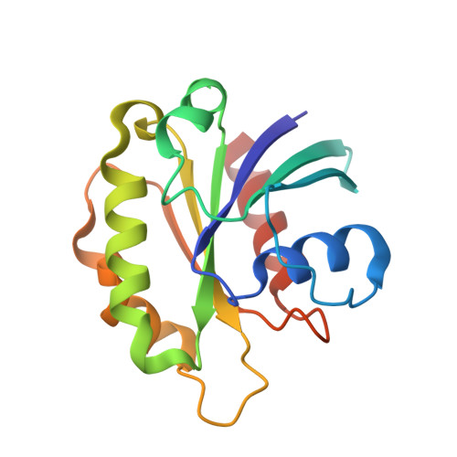
ADP-ribosylation factor 1
B, D, F, H
165Homo sapiensMutation(s): 1 
Gene Names: ARF1
EC: 3.6.5.2
UniProt & NIH Common Fund Data Resources
Find proteins for P84077 (Homo sapiens)
Explore P84077 
Go to UniProtKB:  P84077
PHAROS:  P84077
GTEx:  ENSG00000143761 
Entity Groups  
Sequence Clusters30% Identity50% Identity70% Identity90% Identity95% Identity100% Identity
UniProt GroupP84077
Sequence Annotations
Expand
  • Reference Sequence
Small Molecules
Ligands 3 Unique
IDChains Name / Formula / InChI Key2D Diagram3D Interactions
NAD (Subject of Investigation/LOI)
Query on NAD

Download Ideal Coordinates CCD File 
I [auth A],
L [auth C],
O [auth E],
R [auth G]
NICOTINAMIDE-ADENINE-DINUCLEOTIDE
C21 H27 N7 O14 P2
BAWFJGJZGIEFAR-NNYOXOHSSA-N
GDP
Query on GDP

Download Ideal Coordinates CCD File 
K [auth B],
N [auth D],
Q [auth F],
T [auth H]
GUANOSINE-5'-DIPHOSPHATE
C10 H15 N5 O11 P2
QGWNDRXFNXRZMB-UUOKFMHZSA-N
MG
Query on MG

Download Ideal Coordinates CCD File 
J [auth B],
M [auth D],
P [auth F],
S [auth H]
MAGNESIUM ION
Mg
JLVVSXFLKOJNIY-UHFFFAOYSA-N
Experimental Data & Validation

Experimental Data

  • Method: X-RAY DIFFRACTION
  • Resolution: 3.29 Å
  • R-Value Free:  0.277 (Depositor), 0.280 (DCC) 
  • R-Value Work:  0.236 (Depositor), 0.240 (DCC) 
  • R-Value Observed: 0.239 (Depositor) 
Space Group: P 1 21 1
Unit Cell:
Length ( Å )Angle ( ˚ )
a = 104.711α = 90
b = 105.576β = 104.8
c = 105.839γ = 90
Software Package:
Software NamePurpose
PHENIXrefinement
XDSdata reduction
XSCALEdata scaling
PHENIXphasing

Structure Validation

View Full Validation Report


Ligand Structure Quality Assessment 

Created with Raphaël 2.3.0Worse 01 BetterLigand structure goodness of fit to experimental dataBest fitted NADClick on this verticalbar to view details

Entry History 

Deposition Data

Revision History  (Full details and data files)

  • Version 1.0: 2021-12-15
    Type: Initial release
  • Version 1.1: 2022-12-28
    Changes: Database references
  • Version 1.2: 2024-05-29
    Changes: Data collection