7DIE | pdb_00007die

Crystal structure of M. penetrans Ferritin


Experimental Data Snapshot

  • Method: X-RAY DIFFRACTION
  • Resolution: 1.90 Å
  • R-Value Free: 
    0.177 (Depositor), 0.178 (DCC) 
  • R-Value Work: 
    0.153 (Depositor), 0.154 (DCC) 
  • R-Value Observed: 
    0.154 (Depositor) 

wwPDB Validation 3D Report Full Report

Validation slider image for 7DIE

This is version 1.1 of the entry. See complete history

Literature

Ferritin with Atypical Ferroxidase Centers Takes B-Channels as the Pathway for Fe 2+ Uptake from Mycoplasma .

Wang, W.Zhang, Y.Zhao, G.Wang, H.

(2021) Inorg Chem 60: 7207-7216

  • DOI: https://doi.org/10.1021/acs.inorgchem.1c00265
  • Primary Citation Related Structures: 
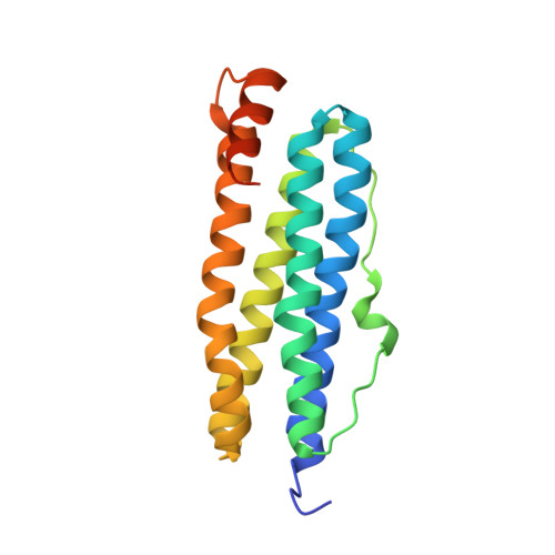
    7DIE

  • PubMed Abstract: 

    Here, we present a 1.9 Å resolution crystal structure of Mycoplasma Penetrans ferritin, which reveals that its ferroxidase center is located on the inner surface of ferritin but not buried within the four-helix of each subunit. Such a ferroxidase center exhibits a lower iron oxidation activity as compared to the reported ferritin. More importantly, we found that Fe 2+ enters into the center via the rarely reported B-channels rather than the normal 3- or 4-fold channels. All these findings may provide the structural bases to explore the new iron oxidation mechanism adopted by this special ferritin, which is beneficial for understanding the relationship between the structure and function of ferritin.


  • Organizational Affiliation
    • Institute of Molecular Science, Shanxi University, Taiyuan 030006, China.

Macromolecule Content 

  • Total Structure Weight: 125.99 kDa 
  • Atom Count: 8,877 
  • Modeled Residue Count: 984 
  • Deposited Residue Count: 1,074 
  • Unique protein chains: 1

Macromolecules

Find similar proteins by:|  3D Structure
Entity ID: 1
MoleculeChains  Sequence LengthOrganismDetailsImage
Ferritin
A, B, C, D, E
A, B, C, D, E, F
179Malacoplasma penetrans HF-2Mutation(s): 0 
Gene Names: MYPE2930
EC: 1.16.3.2
UniProt
Find proteins for Q8EWB1 (Malacoplasma penetrans (strain HF-2))
Explore Q8EWB1 
Go to UniProtKB:  Q8EWB1
Entity Groups
Sequence Clusters30% Identity50% Identity70% Identity90% Identity95% Identity100% Identity
UniProt GroupQ8EWB1
Sequence Annotations
Expand
Reference Sequence

Small Molecules

Ligands 1 Unique
IDChains Name / Formula / InChI Key2D Diagram3D Interactions
FE
(Subject of Investigation/LOI)

Query on FE


Download:Ideal Coordinates CCD File
AA [auth D]
BA [auth E]
CA [auth E]
DA [auth E]
EA [auth E]
AA [auth D],
BA [auth E],
CA [auth E],
DA [auth E],
EA [auth E],
FA [auth E],
G [auth A],
GA [auth E],
H [auth A],
HA [auth F],
I [auth A],
IA [auth F],
J [auth A],
JA [auth F],
K [auth A],
KA [auth F],
L [auth A],
LA [auth F],
M [auth A],
MA [auth F],
N [auth A],
NA [auth F],
O [auth B],
OA [auth F],
P [auth B],
PA [auth F],
Q [auth B],
R [auth B],
S [auth B],
T [auth B],
U [auth C],
V [auth C],
W [auth C],
X [auth C],
Y [auth D],
Z [auth D]
FE (III) ION
Fe
VTLYFUHAOXGGBS-UHFFFAOYSA-N

Experimental Data & Validation

Experimental Data

  • Method: X-RAY DIFFRACTION
  • Resolution: 1.90 Å
  • R-Value Free:  0.177 (Depositor), 0.178 (DCC) 
  • R-Value Work:  0.153 (Depositor), 0.154 (DCC) 
  • R-Value Observed: 0.154 (Depositor) 
Space Group: I 4
Unit Cell:
Length ( Å )Angle ( ˚ )
a = 151.334α = 90
b = 151.334β = 90
c = 125.213γ = 90
Software Package:
Software NamePurpose
PHENIXrefinement
PDB_EXTRACTdata extraction
HKL-3000data reduction
HKL-3000data scaling
AutoSolphasing

Structure Validation

View Full Validation Report


Entry History 

& Funding Information

Deposition Data


Funding OrganizationLocationGrant Number
National Natural Science Foundation of China (NSFC)China21601112, 21671125

Revision History  (Full details and data files)

  • Version 1.0: 2021-05-26
    Type: Initial release
  • Version 1.1: 2024-03-27
    Changes: Data collection, Database references