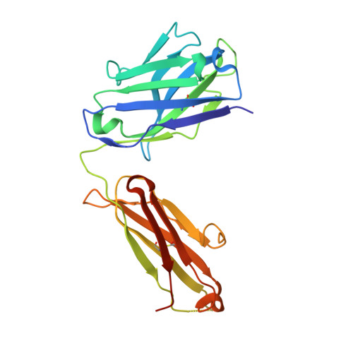
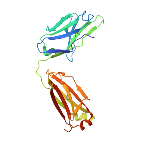
7D85

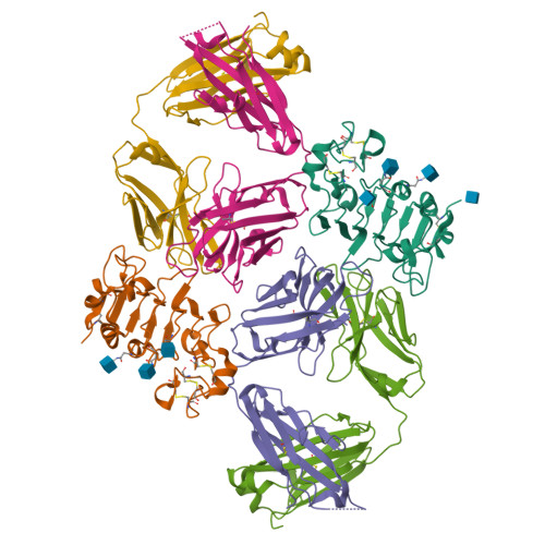
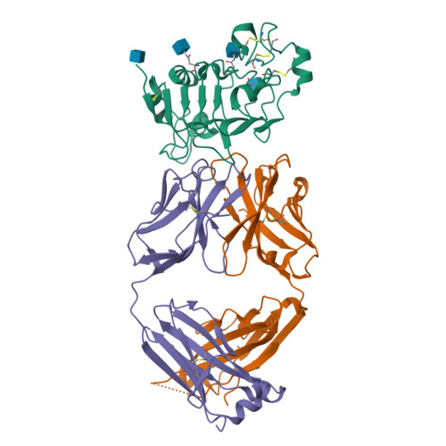
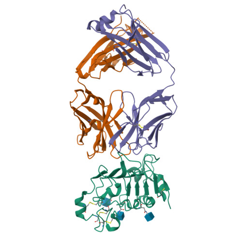
Crystal structure of anti-ErbB3 Fab ISU104 in complex with human ErbB3 extracellular domain 3


Experimental Data Snapshot

  • Method: X-RAY DIFFRACTION
  • Resolution: 2.50 Å
  • R-Value Free: 0.258 
  • R-Value Work: 0.203 
  • R-Value Observed: 0.204 

wwPDB Validation   3D Report Full Report


Ligand Structure Quality Assessment 

Created with Raphaël 2.3.0Worse 01 BetterLigand structure goodness of fit to experimental dataBest fitted NAGClick on this verticalbar to view details

This is version 1.3 of the entry. See complete history


Literature

A Novel Therapeutic Anti-ErbB3, ISU104 Exhibits Potent Antitumorigenic Activity by Inhibiting Ligand Binding and ErbB3 Heterodimerization.

Hong, M.Yoo, Y.Kim, M.Kim, J.Y.Cha, J.S.Choi, M.K.Kim, U.Kim, K.Sohn, Y.Bae, D.Cho, H.S.Hong, S.B.

(2021) Mol Cancer Ther 20: 1142-1152

  • DOI: https://doi.org/10.1158/1535-7163.MCT-20-0907
  • Primary Citation of Related Structures:  
    7D85

  • PubMed Abstract: 

    ErbB3, a member of the ErbB receptor family, is a potent mediator in the development and progression of cancer, and its activation plays pivotal roles in acquired resistance against anti-EGFR therapies and other standard-of-care therapies. Upon ligand (NRG1) binding, ErbB3 forms heterodimers with other ErbB proteins (i.e., EGFR and ErbB2), which allows activation of downstream PI3K/Akt signaling. In this study, we developed a fully human anti-ErbB3 antibody, named ISU104, as an anticancer agent. ISU104 binds potently and specifically to the domain 3 of ErbB3. The complex structure of ErbB3-domain 3::ISU104-Fab revealed that ISU104 binds to the NRG1 binding region of domain 3. The elucidated structure suggested that the binding of ISU104 to ErbB3 would hinder not only ligand binding but also the structural changes required for heterodimerization. Biochemical studies confirmed these predictions. ISU104 inhibited ligand binding, ligand-dependent heterodimerization and phosphorylation, and induced the internalization of ErbB3. As a result, downstream Akt phosphorylation and cell proliferation were inhibited. The anticancer efficacy of ISU104 was demonstrated in xenograft models of various cancers. In summary, a highly potent ErbB3 targeting antibody, ISU104, is suitable for clinical development.


  • Organizational Affiliation

    Research Institute, ISU ABXIS Co., Ltd., Sungnam-si, Republic of Korea (South).


Macromolecules
Find similar proteins by:  (by identity cutoff)  |  3D Structure
Entity ID: 1
MoleculeChains Sequence LengthOrganismDetailsImage
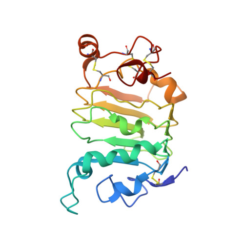
Receptor tyrosine-protein kinase erbB-3
A, D
194Homo sapiensMutation(s): 0 
Gene Names: ERBB3HER3
EC: 2.7.10.1
UniProt & NIH Common Fund Data Resources
Find proteins for P21860 (Homo sapiens)
Explore P21860 
Go to UniProtKB:  P21860
PHAROS:  P21860
GTEx:  ENSG00000065361 
Entity Groups  
Sequence Clusters30% Identity50% Identity70% Identity90% Identity95% Identity100% Identity
UniProt GroupP21860
Glycosylation
Glycosylation Sites: 3Go to GlyGen: P21860-1
Sequence Annotations
Expand
  • Reference Sequence
Find similar proteins by:  (by identity cutoff)  |  3D Structure
Entity ID: 2
MoleculeChains Sequence LengthOrganismDetailsImage
Anti-ErbB3 Fab heavy chain
B, E
228Homo sapiensMutation(s): 0 
Entity Groups  
Sequence Clusters30% Identity50% Identity70% Identity90% Identity95% Identity100% Identity
Sequence Annotations
Expand
  • Reference Sequence
Find similar proteins by:  (by identity cutoff)  |  3D Structure
Entity ID: 3
MoleculeChains Sequence LengthOrganismDetailsImage
Anti-ErbB3 Fab light chain
C, F
216Homo sapiensMutation(s): 0 
Entity Groups  
Sequence Clusters30% Identity50% Identity70% Identity90% Identity95% Identity100% Identity
Sequence Annotations
Expand
  • Reference Sequence
Experimental Data & Validation

Experimental Data

  • Method: X-RAY DIFFRACTION
  • Resolution: 2.50 Å
  • R-Value Free: 0.258 
  • R-Value Work: 0.203 
  • R-Value Observed: 0.204 
  • Space Group: P 1 21 1
Unit Cell:
Length ( Å )Angle ( ˚ )
a = 92.29α = 90
b = 77.78β = 113.41
c = 93.94γ = 90
Software Package:
Software NamePurpose
PHENIXrefinement
XDSdata reduction
XDSdata scaling
PHASERphasing

Structure Validation

View Full Validation Report


Ligand Structure Quality Assessment 

Created with Raphaël 2.3.0Worse 01 BetterLigand structure goodness of fit to experimental dataBest fitted NAGClick on this verticalbar to view details

Entry History 

Deposition Data

Revision History  (Full details and data files)

  • Version 1.0: 2021-04-07
    Type: Initial release
  • Version 1.1: 2021-04-14
    Changes: Database references
  • Version 1.2: 2021-06-16
    Changes: Database references
  • Version 1.3: 2024-10-30
    Changes: Data collection, Database references, Structure summary