7D83 | pdb_00007d83

Crystal structure of HIV-1 integrase catalytic core domain in complex with 2-(tert-butoxy)-2-(2-(3-cyclohexylureido)-3,6-dimethyl-5-(5-methylchroman-6-yl)pyridin-4-yl)acetic acid


Experimental Data Snapshot

  • Method: X-RAY DIFFRACTION
  • Resolution: 2.43 Å
  • R-Value Free: 
    0.278 (Depositor), 0.231 (DCC) 
  • R-Value Work: 
    0.224 (Depositor), 0.226 (DCC) 
  • R-Value Observed: 
    0.226 (Depositor) 

Starting Model: experimental
View more details

wwPDB Validation 3D Report Full Report

Validation slider image for 7D83

Ligand Structure Quality Assessment 


This is version 1.2 of the entry. See complete history

Literature

Discovery of novel HIV-1 integrase-LEDGF/p75 allosteric inhibitors based on a pyridine scaffold forming an intramolecular hydrogen bond.

Sugiyama, S.Akiyama, T.Taoda, Y.Iwaki, T.Matsuoka, E.Akihisa, E.Seki, T.Yoshinaga, T.Kawasuji, T.

(2020) Bioorg Med Chem Lett 33: 127742-127742

  • DOI: https://doi.org/10.1016/j.bmcl.2020.127742
  • Primary Citation Related Structures: 
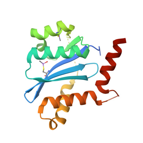
    7D83

  • PubMed Abstract: 

    We have discovered HIV-1 novel integrase-LEDGF/p75 allosteric inhibitors (INLAIs) based on a pyridine scaffold forming an intramolecular hydrogen bond. Scaffolds containing a pyridine moiety have been studied extensively and we have already reported that substituents extending from the C1 position contributed to the antiviral potency. In this study, we designed a new pyridine scaffold 2 with a substituent at the C1 position. Interestingly, during attempts at optimization, we found that the direction of the C1 substituents with an intramolecular hydrogen bond contributed to the antiviral potency. Compound 34f exhibited better antiviral potency against WT and the T174I mutant (EC 50 (WT) = 6.6 nM, EC 50 (T174I) = 270 nM) than BI 224436 (EC 50 (WT) = 22 nM, EC 50 (T174I) > 5000 nM).


  • Organizational Affiliation
    • Shionogi Pharmaceutical Research Center, Shionogi & Co., Ltd., 3-1-1, Futabacho, Toyonaka, Osaka 561-0825, Japan. Electronic address: shuichi.sugiyama@shionogi.co.jp.

Macromolecule Content 

  • Total Structure Weight: 19.25 kDa 
  • Atom Count: 1,156 
  • Modeled Residue Count: 139 
  • Deposited Residue Count: 166 
  • Unique protein chains: 1

Macromolecules

Find similar proteins by:|  3D Structure
Entity ID: 1
MoleculeChains  Sequence LengthOrganismDetailsImage
Integrase166Human immunodeficiency virus type 1 (NEW YORK-5 ISOLATE)Mutation(s): 1 
EC: 2.7.7
UniProt
Find proteins for P12497 (Human immunodeficiency virus type 1 group M subtype B (isolate NY5))
Explore P12497 
Go to UniProtKB:  P12497
Entity Groups
Sequence Clusters30% Identity50% Identity70% Identity90% Identity95% Identity100% Identity
UniProt GroupP12497
Sequence Annotations
Expand
Reference Sequence

Small Molecules

Modified Residues  1 Unique
IDChains TypeFormula2D DiagramParent
CAF
Query on CAF
A
L-PEPTIDE LINKINGC5 H12 As N O3 SCYS

Experimental Data & Validation

Experimental Data

  • Method: X-RAY DIFFRACTION
  • Resolution: 2.43 Å
  • R-Value Free:  0.278 (Depositor), 0.231 (DCC) 
  • R-Value Work:  0.224 (Depositor), 0.226 (DCC) 
  • R-Value Observed: 0.226 (Depositor) 
Space Group: P 31 2 1
Unit Cell:
Length ( Å )Angle ( ˚ )
a = 72.312α = 90
b = 72.312β = 90
c = 65.845γ = 120
Software Package:
Software NamePurpose
HKL-2000data scaling
REFMACrefinement
PDB_EXTRACTdata extraction
HKL-2000data reduction
MOLREPphasing

Structure Validation

View Full Validation Report


Ligand Structure Quality Assessment 


Entry History 

Deposition Data

Revision History  (Full details and data files)

  • Version 1.0: 2021-01-13
    Type: Initial release
  • Version 1.1: 2023-11-29
    Changes: Data collection, Database references, Refinement description
  • Version 1.2: 2024-10-23
    Changes: Structure summary