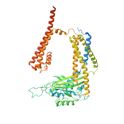
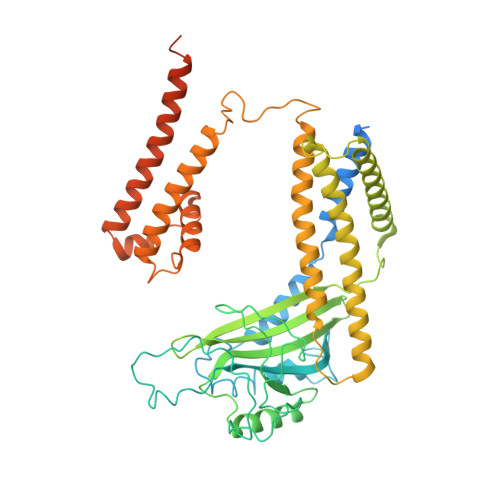
7D7E

Structure of PKD1L3-CTD/PKD2L1 in apo state


Experimental Data Snapshot

  • Method: ELECTRON MICROSCOPY
  • Resolution: 3.40 Å
  • Aggregation State: PARTICLE 
  • Reconstruction Method: SINGLE PARTICLE 

wwPDB Validation   3D Report Full Report


This is version 1.0 of the entry. See complete history


Literature

Structural basis for Ca 2+ activation of the heteromeric PKD1L3/PKD2L1 channel.

Su, Q.Chen, M.Wang, Y.Li, B.Jing, D.Zhan, X.Yu, Y.Shi, Y.

(2021) Nat Commun 12: 4871-4871

  • DOI: https://doi.org/10.1038/s41467-021-25216-z
  • Primary Citation of Related Structures:  
    7D7E, 7D7F

  • PubMed Abstract: 

    The heteromeric complex between PKD1L3, a member of the polycystic kidney disease (PKD) protein family, and PKD2L1, also known as TRPP2 or TRPP3, has been a prototype for mechanistic characterization of heterotetrametric TRP-like channels. Here we show that a truncated PKD1L3/PKD2L1 complex with the C-terminal TRP-fold fragment of PKD1L3 retains both Ca 2+ and acid-induced channel activities. Cryo-EM structures of this core heterocomplex with or without supplemented Ca 2+ were determined at resolutions of 3.1 Å and 3.4 Å, respectively. The heterotetramer, with a pseudo-symmetric TRP architecture of 1:3 stoichiometry, has an asymmetric selectivity filter (SF) guarded by Lys2069 from PKD1L3 and Asp523 from the three PKD2L1 subunits. Ca 2+ -entrance to the SF vestibule is accompanied by a swing motion of Lys2069 on PKD1L3. The S6 of PKD1L3 is pushed inward by the S4-S5 linker of the nearby PKD2L1 (PKD2L1-III), resulting in an elongated intracellular gate which seals the pore domain. Comparison of the apo and Ca 2+ -loaded complexes unveils an unprecedented Ca 2+ binding site in the extracellular cleft of the voltage-sensing domain (VSD) of PKD2L1-III, but not the other three VSDs. Structure-guided mutagenic studies support this unconventional site to be responsible for Ca 2+ -induced channel activation through an allosteric mechanism.


  • Organizational Affiliation

    Key Laboratory of Structural Biology of Zhejiang Province, School of Life Sciences, Westlake University, Hangzhou, Zhejiang Province, China. suqiang@westlake.edu.cn.


Macromolecules
Find similar proteins by:  (by identity cutoff)  |  3D Structure
Entity ID: 1
MoleculeChains Sequence LengthOrganismDetailsImage
Polycystic kidney disease 2-like 1 proteinA [auth B],
B [auth C],
C [auth D]
604Mus musculusMutation(s): 0 
Gene Names: Pkd2l1Trpp3
Membrane Entity: Yes 
UniProt
Find proteins for A2A259 (Mus musculus)
Explore A2A259 
Go to UniProtKB:  A2A259
Entity Groups  
Sequence Clusters30% Identity50% Identity70% Identity90% Identity95% Identity100% Identity
UniProt GroupA2A259
Glycosylation
Glycosylation Sites: 3Go to GlyGen: A2A259-1
Sequence Annotations
Expand
  • Reference Sequence
Find similar proteins by:  (by identity cutoff)  |  3D Structure
Entity ID: 2
MoleculeChains Sequence LengthOrganismDetailsImage
Polycystic kidney disease protein 1-like 3D [auth A]551Mus musculusMutation(s): 0 
Gene Names: Pkd1l371B10
Membrane Entity: Yes 
UniProt
Find proteins for Q2EG98 (Mus musculus)
Explore Q2EG98 
Go to UniProtKB:  Q2EG98
Entity Groups  
Sequence Clusters30% Identity50% Identity70% Identity90% Identity95% Identity100% Identity
UniProt GroupQ2EG98
Glycosylation
Glycosylation Sites: 2Go to GlyGen: Q2EG98-1
Sequence Annotations
Expand
  • Reference Sequence
Small Molecules
Ligands 2 Unique
IDChains Name / Formula / InChI Key2D Diagram3D Interactions
NAG
Query on NAG

Download Ideal Coordinates CCD File 
E [auth B]
F [auth B]
G [auth B]
I [auth C]
J [auth C]
E [auth B],
F [auth B],
G [auth B],
I [auth C],
J [auth C],
K [auth C],
M [auth D],
N [auth D],
P [auth A],
Q [auth A]
2-acetamido-2-deoxy-beta-D-glucopyranose
C8 H15 N O6
OVRNDRQMDRJTHS-FMDGEEDCSA-N
CA
Query on CA

Download Ideal Coordinates CCD File 
H [auth B],
L [auth C],
O [auth D]
CALCIUM ION
Ca
BHPQYMZQTOCNFJ-UHFFFAOYSA-N
Experimental Data & Validation

Experimental Data

  • Method: ELECTRON MICROSCOPY
  • Resolution: 3.40 Å
  • Aggregation State: PARTICLE 
  • Reconstruction Method: SINGLE PARTICLE 

Structure Validation

View Full Validation Report


Entry History & Funding Information

Deposition Data


Funding OrganizationLocationGrant Number
National Natural Science Foundation of China (NSFC)China31930059
National Natural Science Foundation of China (NSFC)China81920108015

Revision History  (Full details and data files)

  • Version 1.0: 2021-08-25
    Type: Initial release