7D6P | pdb_00007d6p

X-ray structure of the intermolecular complex of Clostridium perfringens sortase C with the C-terminal cell wall sorting signal motif.


Experimental Data Snapshot

  • Method: X-RAY DIFFRACTION
  • Resolution: 2.38 Å
  • R-Value Free: 
    0.276 (Depositor), 0.278 (DCC) 
  • R-Value Work: 
    0.244 (Depositor), 0.249 (DCC) 

Starting Model: experimental
View more details

wwPDB Validation 3D Report Full Report

Validation slider image for 7D6P

This is version 1.1 of the entry. See complete history

Literature

X-ray structures of Clostridium perfringens sortase C with C-terminal cell wall sorting motif of LPST demonstrate role of subsite for substrate-binding and structural variations of catalytic site.

Tamai, E.Sekiya, H.Nariya, H.Katayama, S.Kamitori, S.

(2021) Biochem Biophys Res Commun 554: 138-144

  • DOI: https://doi.org/10.1016/j.bbrc.2021.03.106
  • Primary Citation Related Structures: 
    7D6P, 7D6T

  • PubMed Abstract: 

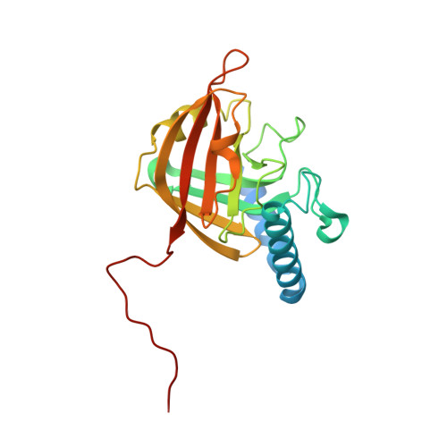
    Pili of Gram-positive bacteria are flexible rod proteins covalently attached to the bacterial cell wall, that play important roles in the initial adhesion of bacterial cells to host tissues and bacterial colonization. Pili are formed by the polymerization of major and minor pilins, catalyzed by class C sortase (SrtC), a family of cysteine transpeptidases. The Gram-positive bacterium Clostridium perfringens has a major pilin (CppA), a minor pilin (CppB), and SrtC (CpSrtC). CpSrtC recognizes the C-terminal cell wall sorting signal motifs with five amino acid residues, LPSTG of CppA and LPETG of CppB, for the polymerization of pili. Here, we report biochemical analysis to detect the formation of Clostridium perfringens pili in vivo, and the X-ray structure of a novel intermolecular substrate-enzyme complex of CpSrtC with a sequence of LPST at the C-terminal site. The results showed that CpSrtC has a subsite for substrate-binding to aid polymerization of pili, and that the catalytic site has structural variations, giving insights into the enzyme catalytic reaction mechanism and affinities for the C-terminal cell wall sorting signal motif sequences.


  • Organizational Affiliation
    • Department of Infectious Disease, College of Pharmaceutical Sciences, Matsuyama University, 4-2 Bunkyo-cho, Matsuyama, Ehime, 790-8578, Japan; Life Science Research Center and Faculty of Medicine, Kagawa University, 1750-1 Ikenobe, Miki-cho, Kita-gun, Kagawa, 761-0793, Japan.

Macromolecule Content 

  • Total Structure Weight: 25.02 kDa 
  • Atom Count: 1,580 
  • Modeled Residue Count: 198 
  • Deposited Residue Count: 220 
  • Unique protein chains: 1

Macromolecules

Find similar proteins by:|  3D Structure
Entity ID: 1
MoleculeChains  Sequence LengthOrganismDetailsImage
Sortase family protein220Clostridium perfringens SM101Mutation(s): 0 
Gene Names: CPR_0146
UniProt
Find proteins for Q0SWL7 (Clostridium perfringens (strain SM101 / Type A))
Explore Q0SWL7 
Go to UniProtKB:  Q0SWL7
Entity Groups
Sequence Clusters30% Identity50% Identity70% Identity90% Identity95% Identity100% Identity
UniProt GroupQ0SWL7
Sequence Annotations
Expand
Reference Sequence

Experimental Data & Validation

Experimental Data

  • Method: X-RAY DIFFRACTION
  • Resolution: 2.38 Å
  • R-Value Free:  0.276 (Depositor), 0.278 (DCC) 
  • R-Value Work:  0.244 (Depositor), 0.249 (DCC) 
Space Group: P 32 2 1
Unit Cell:
Length ( Å )Angle ( ˚ )
a = 104.21α = 90
b = 104.21β = 90
c = 44.87γ = 120
Software Package:
Software NamePurpose
REFMACrefinement
XDSdata reduction
XDSdata scaling
MOLREPphasing

Structure Validation

View Full Validation Report


Entry History 

& Funding Information

Deposition Data


Funding OrganizationLocationGrant Number
Japan Society for the Promotion of Science (JSPS)Japan18K06087

Revision History  (Full details and data files)

  • Version 1.0: 2021-06-02
    Type: Initial release
  • Version 1.1: 2023-11-29
    Changes: Data collection, Database references, Derived calculations, Refinement description