7D3E

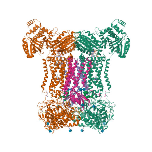
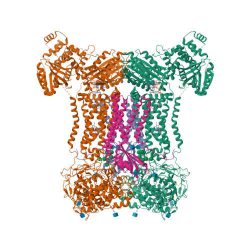
Cryo-EM structure of human DUOX1-DUOXA1 in low-calcium state


Experimental Data Snapshot

  • Method: ELECTRON MICROSCOPY
  • Resolution: 2.80 Å
  • Aggregation State: PARTICLE 
  • Reconstruction Method: SINGLE PARTICLE 

wwPDB Validation   3D Report Full Report


This is version 1.2 of the entry. See complete history


Literature

Structures of human dual oxidase 1 complex in low-calcium and high-calcium states.

Wu, J.X.Liu, R.Song, K.Chen, L.

(2021) Nat Commun 12: 155-155

  • DOI: https://doi.org/10.1038/s41467-020-20466-9
  • Primary Citation of Related Structures:  
    7D3E, 7D3F

  • PubMed Abstract: 

    Dual oxidases (DUOXs) produce hydrogen peroxide by transferring electrons from intracellular NADPH to extracellular oxygen. They are involved in many crucial biological processes and human diseases, especially in thyroid diseases. DUOXs are protein complexes co-assembled from the catalytic DUOX subunits and the auxiliary DUOXA subunits and their activities are regulated by intracellular calcium concentrations. Here, we report the cryo-EM structures of human DUOX1-DUOXA1 complex in both high-calcium and low-calcium states. These structures reveal the DUOX1 complex is a symmetric 2:2 hetero-tetramer stabilized by extensive inter-subunit interactions. Substrate NADPH and cofactor FAD are sandwiched between transmembrane domain and the cytosolic dehydrogenase domain of DUOX. In the presence of calcium ions, intracellular EF-hand modules might enhance the catalytic activity of DUOX by stabilizing the dehydrogenase domain in a conformation that allows electron transfer.


  • Organizational Affiliation

    State Key Laboratory of Membrane Biology, College of Future Technology, Institute of Molecular Medicine, Beijing Key Laboratory of Cardiometabolic Molecular Medicine, Peking University, 100871, Beijing, China.


Macromolecules
Find similar proteins by:  (by identity cutoff)  |  3D Structure
Entity ID: 1
MoleculeChains Sequence LengthOrganismDetailsImage
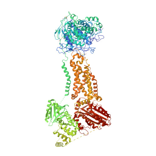
Dual oxidase 1
A, C
1,551Homo sapiensMutation(s): 1 
Gene Names: DUOX1DUOXLNOX1THOX1
EC: 1.11.1 (PDB Primary Data), 1.6.3.1 (PDB Primary Data)
Membrane Entity: Yes 
UniProt & NIH Common Fund Data Resources
Find proteins for Q9NRD9 (Homo sapiens)
Explore Q9NRD9 
Go to UniProtKB:  Q9NRD9
PHAROS:  Q9NRD9
GTEx:  ENSG00000137857 
Entity Groups  
Sequence Clusters30% Identity50% Identity70% Identity90% Identity95% Identity100% Identity
UniProt GroupQ9NRD9
Glycosylation
Glycosylation Sites: 3Go to GlyGen: Q9NRD9-1
Sequence Annotations
Expand
  • Reference Sequence
Find similar proteins by:  (by identity cutoff)  |  3D Structure
Entity ID: 2
MoleculeChains Sequence LengthOrganismDetailsImage
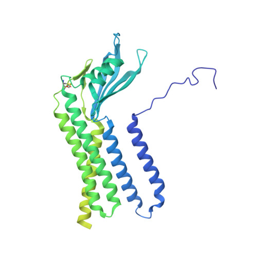
Isoform 2 of Dual oxidase maturation factor 1
B, D
483Homo sapiensMutation(s): 0 
Gene Names: DUOXA1NIPNUMBIP
Membrane Entity: Yes 
UniProt & NIH Common Fund Data Resources
Find proteins for Q1HG43 (Homo sapiens)
Explore Q1HG43 
Go to UniProtKB:  Q1HG43
PHAROS:  Q1HG43
GTEx:  ENSG00000140254 
Entity Groups  
Sequence Clusters30% Identity50% Identity70% Identity90% Identity95% Identity100% Identity
UniProt GroupQ1HG43
Glycosylation
Glycosylation Sites: 3Go to GlyGen: Q1HG43-1
Sequence Annotations
Expand
  • Reference Sequence
Oligosaccharides

Help

Entity ID: 3
MoleculeChains Length2D Diagram Glycosylation3D Interactions
alpha-D-mannopyranose-(1-2)-alpha-D-mannopyranose-(1-2)-alpha-D-mannopyranose-(1-3)-[alpha-D-mannopyranose-(1-2)-alpha-D-mannopyranose-(1-6)-[alpha-D-mannopyranose-(1-3)]alpha-D-mannopyranose-(1-6)]beta-D-mannopyranose-(1-4)-2-acetamido-2-deoxy-beta-D-glucopyranose-(1-4)-2-acetamido-2-deoxy-beta-D-glucopyranose
E, F
10N-Glycosylation
Glycosylation Resources
GlyTouCan:  G40702WU
GlyCosmos:  G40702WU
GlyGen:  G40702WU
Small Molecules
Ligands 5 Unique
IDChains Name / Formula / InChI Key2D Diagram3D Interactions
FAD (Subject of Investigation/LOI)
Query on FAD

Download Ideal Coordinates CCD File 
J [auth A],
U [auth C]
FLAVIN-ADENINE DINUCLEOTIDE
C27 H33 N9 O15 P2
VWWQXMAJTJZDQX-UYBVJOGSSA-N
NDP (Subject of Investigation/LOI)
Query on NDP

Download Ideal Coordinates CCD File 
I [auth A],
T [auth C]
NADPH DIHYDRO-NICOTINAMIDE-ADENINE-DINUCLEOTIDE PHOSPHATE
C21 H30 N7 O17 P3
ACFIXJIJDZMPPO-NNYOXOHSSA-N
HEM (Subject of Investigation/LOI)
Query on HEM

Download Ideal Coordinates CCD File 
G [auth A],
H [auth A],
R [auth C],
S [auth C]
PROTOPORPHYRIN IX CONTAINING FE
C34 H32 Fe N4 O4
KABFMIBPWCXCRK-RGGAHWMASA-L
NAG
Query on NAG

Download Ideal Coordinates CCD File 
AA [auth D]
BA [auth D]
K [auth A]
L [auth A]
M [auth A]
AA [auth D],
BA [auth D],
K [auth A],
L [auth A],
M [auth A],
P [auth B],
Q [auth B],
V [auth C],
W [auth C],
X [auth C]
2-acetamido-2-deoxy-beta-D-glucopyranose
C8 H15 N O6
OVRNDRQMDRJTHS-FMDGEEDCSA-N
NA
Query on NA

Download Ideal Coordinates CCD File 
N [auth A],
O [auth A],
Y [auth C],
Z [auth C]
SODIUM ION
Na
FKNQFGJONOIPTF-UHFFFAOYSA-N
Experimental Data & Validation

Experimental Data

  • Method: ELECTRON MICROSCOPY
  • Resolution: 2.80 Å
  • Aggregation State: PARTICLE 
  • Reconstruction Method: SINGLE PARTICLE 

Structure Validation

View Full Validation Report


Entry History & Funding Information

Deposition Data


Funding OrganizationLocationGrant Number
National Natural Science Foundation of China (NSFC)China91957201
National Natural Science Foundation of China (NSFC)China31622021

Revision History  (Full details and data files)

  • Version 1.0: 2020-12-09
    Type: Initial release
  • Version 1.1: 2021-06-23
    Changes: Database references
  • Version 1.2: 2024-11-06
    Changes: Data collection, Database references, Structure summary