7D2F

Lp major histidine acid phosphatase mutant D281A/5'-AMP


Experimental Data Snapshot

  • Method: X-RAY DIFFRACTION
  • Resolution: 2.60 Å
  • R-Value Free: 0.256 
  • R-Value Work: 0.220 
  • R-Value Observed: 0.222 

Starting Model: experimental
View more details

wwPDB Validation   3D Report Full Report


Ligand Structure Quality Assessment 


This is version 1.2 of the entry. See complete history


Literature

Structural insights into a new substrate binding mode of a histidine acid phosphatase from Legionella pneumophila.

Guo, Y.Zhou, D.Zhang, H.Zhang, N.N.Qi, X.Chen, X.Chen, Q.Li, J.Ge, H.Teng, Y.B.

(2021) Biochem Biophys Res Commun 540: 90-94

  • DOI: https://doi.org/10.1016/j.bbrc.2020.12.070
  • Primary Citation of Related Structures:  
    7D2F, 7DOQ

  • PubMed Abstract: 

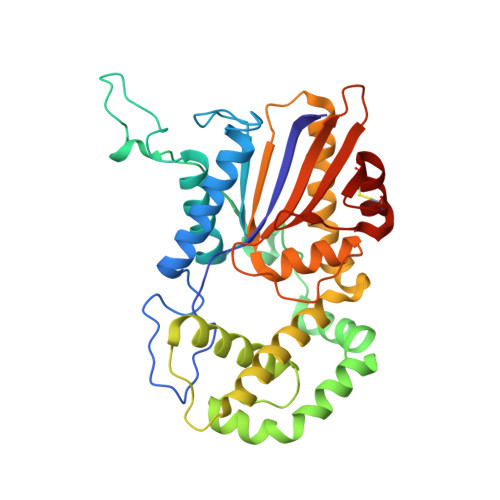
    MapA is a histidine acid phosphatase (HAP) from Legionella pneumophila that catalyzes the hydroxylation of a phosphoryl group from phosphomonoesters by an active-site histidine. Several structures of HAPs, including MapA, in complex with the inhibitor tartrate have been solved and the substrate binding tunnel identified; however, the substrate recognition mechanism remains unknown. To gain insight into the mechanism of substrate recognition, the crystal structures of apo-MapA and the MapA D281A mutant in complex with 5'-AMP were solved at 2.2 and 2.6 Å resolution, respectively. The structure of the MapA D281A /5'-AMP complex reveals that the 5'-AMP fits fully into the substrate binding tunnel, with the 2'-hydroxyl group of the ribose moiety stabilized by Glu201 and the adenine moiety sandwiched between His205 and Phe237. This is the second structure of a HAP/AMP complex solved with 5'-AMP binding in a unique manner in the active site. The structure presents a new substrate recognition mechanism of HAPs.


  • Organizational Affiliation

    School of Life Sciences, Anhui Medical University, Hefei, Anhui, 230032, People's Republic of China. Electronic address: guoyu_joy@163.com.


Macromolecules
Find similar proteins by:  (by identity cutoff)  |  3D Structure
Entity ID: 1
MoleculeChains Sequence LengthOrganismDetailsImage
Major acid phosphatase
A, B
328Legionella pneumophila subsp. pneumophila str. Philadelphia 1Mutation(s): 1 
Gene Names: lpg1119
EC: 3.1.3.2
UniProt
Find proteins for Q5ZWG6 (Legionella pneumophila subsp. pneumophila (strain Philadelphia 1 / ATCC 33152 / DSM 7513))
Explore Q5ZWG6 
Go to UniProtKB:  Q5ZWG6
Entity Groups  
Sequence Clusters30% Identity50% Identity70% Identity90% Identity95% Identity100% Identity
UniProt GroupQ5ZWG6
Sequence Annotations
Expand
  • Reference Sequence
Experimental Data & Validation

Experimental Data

  • Method: X-RAY DIFFRACTION
  • Resolution: 2.60 Å
  • R-Value Free: 0.256 
  • R-Value Work: 0.220 
  • R-Value Observed: 0.222 
  • Space Group: P 21 21 21
Unit Cell:
Length ( Å )Angle ( ˚ )
a = 55.61α = 90
b = 118.41β = 90
c = 118.8γ = 90
Software Package:
Software NamePurpose
REFMACrefinement
HKL-2000data scaling
PDB_EXTRACTdata extraction
HKL-2000data reduction
PHENIXphasing

Structure Validation

View Full Validation Report


Ligand Structure Quality Assessment 


Entry History & Funding Information

Deposition Data


Funding OrganizationLocationGrant Number
National Natural Science Foundation of China (NSFC)China31700650

Revision History  (Full details and data files)

  • Version 1.0: 2021-09-22
    Type: Initial release
  • Version 1.1: 2022-07-06
    Changes: Database references
  • Version 1.2: 2023-11-29
    Changes: Data collection, Refinement description