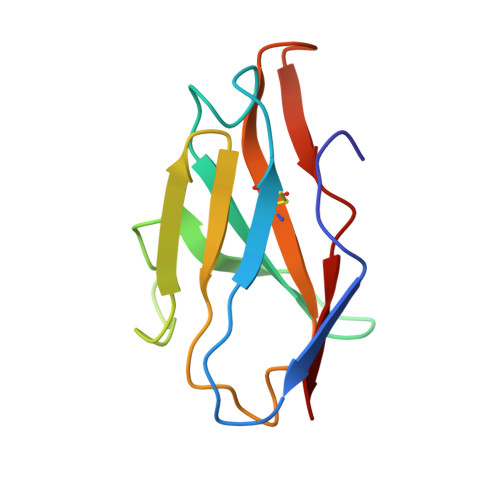
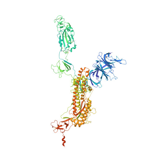
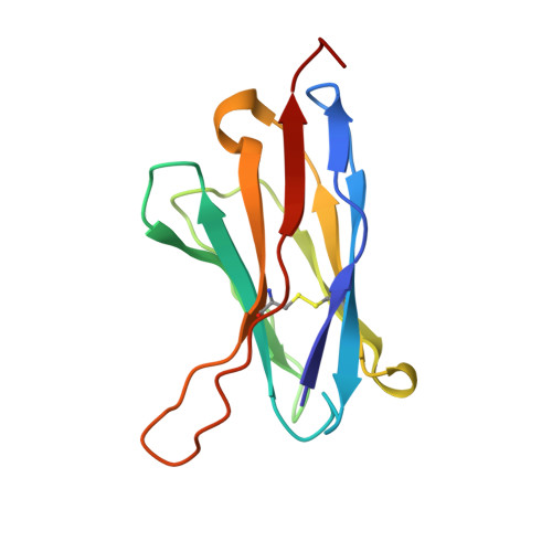
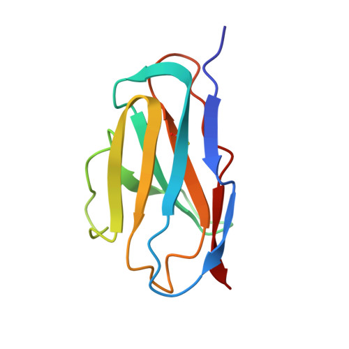
Primary Citation Related Structures: 7CWS, 7CWT, 7CWU
Organizational Affiliation:
CAS Key Laboratory of Infection and Immunity, National Laboratory of Macromolecules, Institute of Biophysics, Chinese Academy of Sciences, Beijing, 100101, China.
University of Chinese Academy of Sciences, Beijing, 100049, China.
Department of Respiratory and Critical Care Medicine, West China Medical School/West China Hospital, Sichuan University, Chengdu, Sichuan, 610041, China.
State Key Laboratory of Pathogen and Biosecurity, Beijing Institute of Microbiology and Epidemiology, AMMS, Beijing, 100071, China.
National Health Commission of the People's Republic of China, Key laboratory of Enteric Pathogenic Microbiology (Jiangsu Provincial Center for Disease Control and Prevention), Nanjing, Jiangsu, 210009, China.
Department of Microbiology, Zhejiang Provincial Center for Disease Control and Prevention, Hangzhou, Zhejiang, 310000, China.
Department of Respiratory and Critical Care Medicine, West China Medical School/West China Hospital, Sichuan University, Chengdu, Sichuan, 610041, China. weimi003@scu.edu.cn.
National Health Commission of the People's Republic of China, Key laboratory of Enteric Pathogenic Microbiology (Jiangsu Provincial Center for Disease Control and Prevention), Nanjing, Jiangsu, 210009, China. jszfc@vip.sina.com.
State Key Laboratory of Pathogen and Biosecurity, Beijing Institute of Microbiology and Epidemiology, AMMS, Beijing, 100071, China. qincf@bmi.ac.cn.
CAS Key Laboratory of Infection and Immunity, National Laboratory of Macromolecules, Institute of Biophysics, Chinese Academy of Sciences, Beijing, 100101, China. xiangxi@ibp.ac.cn.
University of Chinese Academy of Sciences, Beijing, 100049, China. xiangxi@ibp.ac.cn.
Guangzhou Regenerative Medicine and Health Guangdong Laboratory, Guangzhou, Guangdong, 510200, China. xiangxi@ibp.ac.cn.
EA [auth B] FA [auth B] GA [auth B] HA [auth B] IA [auth B]
EA [auth B], FA [auth B], GA [auth B], HA [auth B], IA [auth B], JA [auth C], KA [auth C], LA [auth C], MA [auth C], NA [auth C], OA [auth C], PA [auth A], QA [auth A], RA [auth A], SA [auth A], TA [auth A]
2-acetamido-2-deoxy-beta-D-glucopyranose C8 H15 N O6 OVRNDRQMDRJTHS-FMDGEEDCSA-N