7CR7

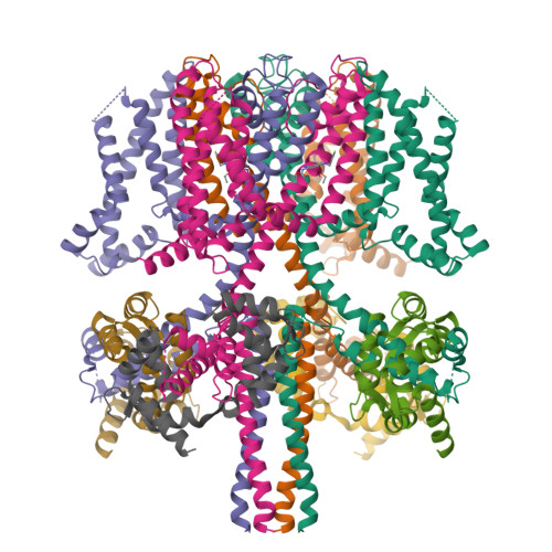
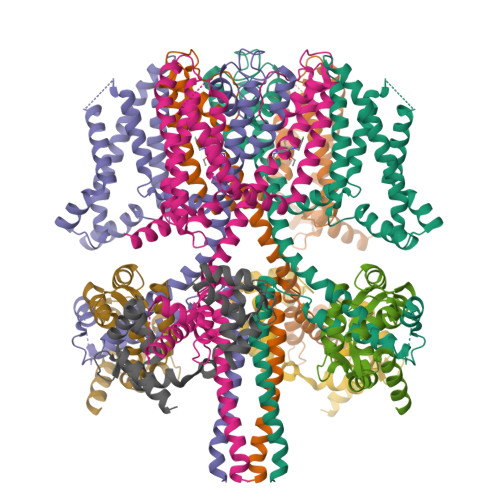
human KCNQ2-CaM in complex with retigabine


Experimental Data Snapshot

  • Method: ELECTRON MICROSCOPY
  • Resolution: 3.70 Å
  • Aggregation State: PARTICLE 
  • Reconstruction Method: SINGLE PARTICLE 

wwPDB Validation   3D Report Full Report


This is version 1.3 of the entry. See complete history


Literature

Molecular basis for ligand activation of the human KCNQ2 channel.

Li, X.Zhang, Q.Guo, P.Fu, J.Mei, L.Lv, D.Wang, J.Lai, D.Ye, S.Yang, H.Guo, J.

(2021) Cell Res 31: 52-61

  • DOI: https://doi.org/10.1038/s41422-020-00410-8
  • Primary Citation of Related Structures:  
    7CR0, 7CR1, 7CR2, 7CR3, 7CR4, 7CR7

  • PubMed Abstract: 

    The voltage-gated potassium channel KCNQ2 is responsible for M-current in neurons and is an important drug target to treat epilepsy, pain and several other diseases related to neuronal hyper-excitability. A list of synthetic compounds have been developed to directly activate KCNQ2, yet our knowledge of their activation mechanism is limited, due to lack of high-resolution structures. Here, we report cryo-electron microscopy (cryo-EM) structures of the human KCNQ2 determined in apo state and in complex with two activators, ztz240 or retigabine, which activate KCNQ2 through different mechanisms. The activator-bound structures, along with electrophysiology analysis, reveal that ztz240 binds at the voltage-sensing domain and directly stabilizes it at the activated state, whereas retigabine binds at the pore domain and activates the channel by an allosteric modulation. By accurately defining ligand-binding sites, these KCNQ2 structures not only reveal different ligand recognition and activation mechanisms, but also provide a structural basis for drug optimization and design.


  • Organizational Affiliation

    Department of Biophysics, and Department of Pathology of Sir Run Run Shaw Hospital, Zhejiang University School of Medicine, Hangzhou, Zhejiang, 310058, China.


Macromolecules
Find similar proteins by:  (by identity cutoff)  |  3D Structure
Entity ID: 1
MoleculeChains Sequence LengthOrganismDetailsImage
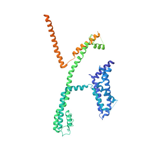
Potassium voltage-gated channel subfamily KQT member 2A,
C [auth B],
E [auth D],
G
656Homo sapiensMutation(s): 0 
Gene Names: KCNQ2
Membrane Entity: Yes 
UniProt & NIH Common Fund Data Resources
Find proteins for O43526 (Homo sapiens)
Explore O43526 
Go to UniProtKB:  O43526
PHAROS:  O43526
GTEx:  ENSG00000075043 
Entity Groups  
Sequence Clusters30% Identity50% Identity70% Identity90% Identity95% Identity100% Identity
UniProt GroupO43526
Sequence Annotations
Expand
  • Reference Sequence
Find similar proteins by:  (by identity cutoff)  |  3D Structure
Entity ID: 2
MoleculeChains Sequence LengthOrganismDetailsImage
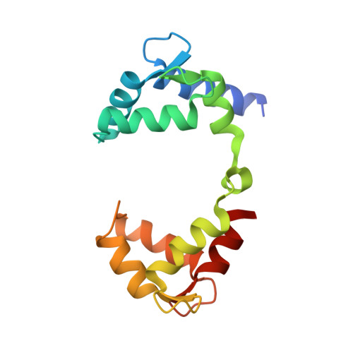
Calmodulin-3B [auth F],
D [auth C],
F [auth E],
H
149Homo sapiensMutation(s): 0 
UniProt & NIH Common Fund Data Resources
Find proteins for P0DP25 (Homo sapiens)
Explore P0DP25 
Go to UniProtKB:  P0DP25
PHAROS:  P0DP25
GTEx:  ENSG00000160014 
Entity Groups  
Sequence Clusters30% Identity50% Identity70% Identity90% Identity95% Identity100% Identity
UniProt GroupP0DP25
Sequence Annotations
Expand
  • Reference Sequence
Experimental Data & Validation

Experimental Data

  • Method: ELECTRON MICROSCOPY
  • Resolution: 3.70 Å
  • Aggregation State: PARTICLE 
  • Reconstruction Method: SINGLE PARTICLE 

Structure Validation

View Full Validation Report


Entry History & Funding Information

Deposition Data


Funding OrganizationLocationGrant Number
Ministry of Science and Technology (MoST, China)China2018YFA0508100
National Natural Science Foundation of China (NSFC)China31870724

Revision History  (Full details and data files)

  • Version 1.0: 2020-09-16
    Type: Initial release
  • Version 1.1: 2020-11-11
    Changes: Structure summary
  • Version 1.2: 2021-01-13
    Changes: Database references
  • Version 1.3: 2024-03-27
    Changes: Data collection, Database references