7CMW | pdb_00007cmw

Complex structure of PARP1 catalytic domain with pamiparib


Experimental Data Snapshot

  • Method: X-RAY DIFFRACTION
  • Resolution: 2.70 Å
  • R-Value Free: 
    0.258 (Depositor), 0.257 (DCC) 
  • R-Value Work: 
    0.204 (Depositor), 0.204 (DCC) 
  • R-Value Observed: 
    0.209 (Depositor) 

Starting Model: experimental
View more details

wwPDB Validation 3D Report Full Report

Validation slider image for 7CMW

Ligand Structure Quality Assessment 


This is version 1.2 of the entry. See complete history

Literature

Discovery of Pamiparib (BGB-290), a Potent and Selective Poly (ADP-ribose) Polymerase (PARP) Inhibitor in Clinical Development.

Wang, H.Ren, B.Liu, Y.Jiang, B.Guo, Y.Wei, M.Luo, L.Kuang, X.Qiu, M.Lv, L.Xu, H.Qi, R.Yan, H.Xu, D.Wang, Z.Huo, C.X.Zhu, Y.Zhao, Y.Wu, Y.Qin, Z.Su, D.Tang, T.Wang, F.Sun, X.Feng, Y.Peng, H.Wang, X.Gao, Y.Liu, Y.Gong, W.Yu, F.Liu, X.Wang, L.Zhou, C.

(2020) J Med Chem 63: 15541-15563

  • DOI: https://doi.org/10.1021/acs.jmedchem.0c01346
  • Primary Citation Related Structures: 
    7CMW

  • PubMed Abstract: 

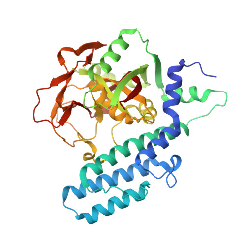
    Poly (ADP-ribose) polymerase (PARP) plays a significant role in DNA repair responses; therefore, this enzyme is targeted by PARP inhibitors in cancer therapy. Here we have developed a number of fused tetra- or pentacyclic dihydrodiazepinoindolone derivatives with excellent PARP enzymatic and cellular PARylation inhibition activities. These efforts led to the identification of pamiparib (BGB-290, 139 ), which displays excellent PARP-1 and PARP-2 inhibition with IC 50 of 1.3 and 0.9 nM, respectively. In a cellular PARylation assay, this compound inhibits PARP activity with IC 50 = 0.2 nM. Cocrystal of pamiparib shows similar binding sites with PARP with other PARP inhibitors, but pamiparib is not a P-gp substrate and shows excellent drug metabolism and pharmacokinetics (DMPK) properties with significant brain penetration (17-19%, mice). The compound is currently being investigated in phase III clinical trials as a maintenance therapy in platinum-sensitive ovarian cancer and gastric cancer.

Macromolecule Content 

  • Total Structure Weight: 80.04 kDa 
  • Atom Count: 5,648 
  • Modeled Residue Count: 700 
  • Deposited Residue Count: 708 
  • Unique protein chains: 1

Macromolecules

Find similar proteins by:|  3D Structure
Entity ID: 1
MoleculeChains  Sequence LengthOrganismDetailsImage
Poly [ADP-ribose] polymerase 1
A, B
354Homo sapiensMutation(s): 1 
Gene Names: PARP1ADPRTPPOL
EC: 2.4.2.30 (PDB Primary Data), 2.4.2 (PDB Primary Data)
UniProt & NIH Common Fund Data Resources
Find proteins for P09874 (Homo sapiens)
Explore P09874 
Go to UniProtKB:  P09874
PHAROS:  P09874
GTEx:  ENSG00000143799 
Entity Groups
Sequence Clusters30% Identity50% Identity70% Identity90% Identity95% Identity100% Identity
UniProt GroupP09874
Sequence Annotations
Expand
Reference Sequence

Experimental Data & Validation

Experimental Data

  • Method: X-RAY DIFFRACTION
  • Resolution: 2.70 Å
  • R-Value Free:  0.258 (Depositor), 0.257 (DCC) 
  • R-Value Work:  0.204 (Depositor), 0.204 (DCC) 
  • R-Value Observed: 0.209 (Depositor) 
Space Group: P 21 21 21
Unit Cell:
Length ( Å )Angle ( ˚ )
a = 47.798α = 90
b = 92.427β = 90
c = 164.495γ = 90
Software Package:
Software NamePurpose
PHENIXrefinement
HKL-2000data reduction
HKL-2000data scaling
PHENIXphasing

Structure Validation

View Full Validation Report


Ligand Structure Quality Assessment 


Entry History 

Deposition Data

Revision History  (Full details and data files)

  • Version 1.0: 2020-12-16
    Type: Initial release
  • Version 1.1: 2021-01-13
    Changes: Database references
  • Version 1.2: 2023-11-29
    Changes: Data collection, Database references, Refinement description