7CM9 | pdb_00007cm9

DMSP lyase DddX


Experimental Data Snapshot

  • Method: X-RAY DIFFRACTION
  • Resolution: 2.25 Å
  • R-Value Free: 
    0.230 (Depositor), 0.223 (DCC) 
  • R-Value Work: 
    0.184 (Depositor), 0.181 (DCC) 
  • R-Value Observed: 
    0.186 (Depositor) 

wwPDB Validation 3D Report Full Report

Validation slider image for 7CM9

Ligand Structure Quality Assessment 


This is version 1.1 of the entry. See complete history

Literature

A novel ATP dependent dimethylsulfoniopropionate lyase in bacteria that releases dimethyl sulfide and acryloyl-CoA.

Li, C.Y.Wang, X.J.Chen, X.L.Sheng, Q.Zhang, S.Wang, P.Quareshy, M.Rihtman, B.Shao, X.Gao, C.Li, F.Li, S.Zhang, W.Zhang, X.H.Yang, G.P.Todd, J.D.Chen, Y.Zhang, Y.Z.

(2021) Elife 10

  • DOI: https://doi.org/10.7554/eLife.64045
  • Primary Citation Related Structures: 
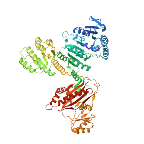
    7CM9

  • PubMed Abstract: 

    Dimethylsulfoniopropionate (DMSP) is an abundant and ubiquitous organosulfur molecule in marine environments with important roles in global sulfur and nutrient cycling. Diverse DMSP lyases in some algae, bacteria, and fungi cleave DMSP to yield gaseous dimethyl sulfide (DMS), an infochemical with important roles in atmospheric chemistry. Here, we identified a novel ATP-dependent DMSP lyase, DddX. DddX belongs to the acyl-CoA synthetase superfamily and is distinct from the eight other known DMSP lyases. DddX catalyses the conversion of DMSP to DMS via a two-step reaction: the ligation of DMSP with CoA to form the intermediate DMSP-CoA, which is then cleaved to DMS and acryloyl-CoA. The novel catalytic mechanism was elucidated by structural and biochemical analyses. DddX is found in several Alphaproteobacteria, Gammaproteobacteria, and Firmicutes, suggesting that this new DMSP lyase may play an overlooked role in DMSP/DMS cycles.


  • Organizational Affiliation
    • State Key Lab of Microbial Technology, Marine Biotechnology Research Center, Shandong University, Qingdao, China.

Macromolecule Content 

  • Total Structure Weight: 329.2 kDa 
  • Atom Count: 22,513 
  • Modeled Residue Count: 2,771 
  • Deposited Residue Count: 2,992 
  • Unique protein chains: 1

Macromolecules

Find similar proteins by:|  3D Structure
Entity ID: 1
MoleculeChains  Sequence LengthOrganismDetailsImage
DMSP lyase
A, B, C, D
748Psychrobacter sp.Mutation(s): 0 

Small Molecules

Ligands 2 Unique
IDChains Name / Formula / InChI Key2D Diagram3D Interactions
ATP
(Subject of Investigation/LOI)

Query on ATP


Download:Ideal Coordinates CCD File
E [auth A],
G [auth B],
I [auth C],
K [auth D]
ADENOSINE-5'-TRIPHOSPHATE
C10 H16 N5 O13 P3
ZKHQWZAMYRWXGA-KQYNXXCUSA-N
SO4

Query on SO4


Download:Ideal Coordinates CCD File
F [auth A],
H [auth B],
J [auth C],
L [auth D]
SULFATE ION
O4 S
QAOWNCQODCNURD-UHFFFAOYSA-L

Experimental Data & Validation

Experimental Data

  • Method: X-RAY DIFFRACTION
  • Resolution: 2.25 Å
  • R-Value Free:  0.230 (Depositor), 0.223 (DCC) 
  • R-Value Work:  0.184 (Depositor), 0.181 (DCC) 
  • R-Value Observed: 0.186 (Depositor) 
Space Group: C 1 2 1
Unit Cell:
Length ( Å )Angle ( ˚ )
a = 143.565α = 90
b = 88.798β = 99.94
c = 273.263γ = 90
Software Package:
Software NamePurpose
PHENIXrefinement
HKL-2000data reduction
HKL-2000data scaling
PDB_EXTRACTdata extraction
PHENIXphasing

Structure Validation

View Full Validation Report


Ligand Structure Quality Assessment 


Entry History 

Deposition Data

Revision History  (Full details and data files)

  • Version 1.0: 2021-05-19
    Type: Initial release
  • Version 1.1: 2024-03-27
    Changes: Data collection, Database references